Plant diseases and their control measures
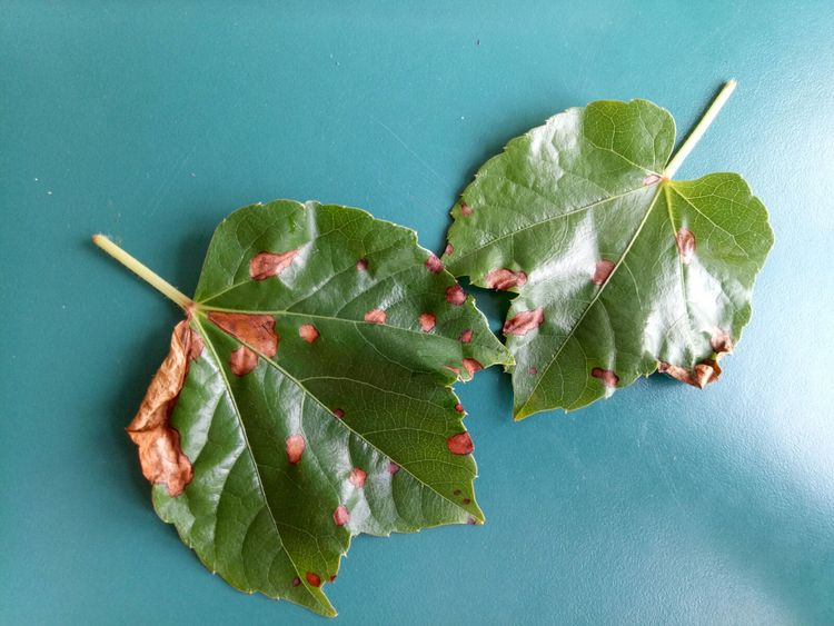
Virginia creeper anthrax
Symptoms:
Leaf lesions are nearly circular or irregular in shape, brown, grayish-brown to grayish-white, with dark brown to blackish-brown edges, a lighter color in the center and slight cloud-like patterns, and scattered small black dots.
Control measures:
① Clean the orchard in winter and spring, removing overly dense shade branches and diseased plant debris, then spray the entire plant once with 1% Bordeaux mixture.
② During the disease period, depending on the weather and the severity of the disease, alternately spray with 30% copper oxychloride suspension, or 40% polysulfide suspension, or 30% mixed sulfur suspension at a dilution of 600-800 times.
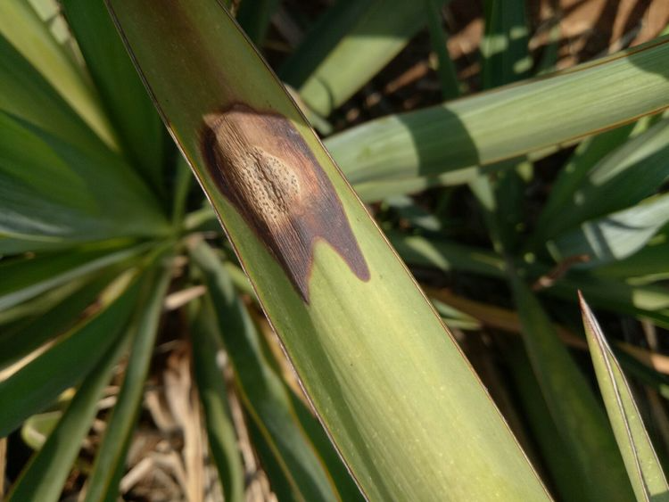
Sisal anthrax
Anthracnose in sisal: [Symptoms] Both sides of the leaves can be infected. Initially, light green to dark brown, slightly sunken lesions appear on the leaf surface, gradually turning blackish-brown. In later stages, the lesions become irregular, with many small black dots scattered on them. When dry, the lesions shrink, and the fibers easily break.
[Disease Name] Sisal Anthracnose
[Pathogen] Colletotrichum agaves Cav., a deuteromycete.
【Prevention and Control Methods】① Maintain good hygiene in the hemp field, cut off diseased leaves and burn them; ② Adopt reasonable plant spacing to avoid overcrowding and ensure good ventilation and light penetration in the hemp field; ③ Strengthen hemp field management before winter and increase potassium fertilizer application to improve the hemp plants' ability to withstand cold and disease; ④ When the disease is severe, 1% Bordeaux mixture or 0.3% to 0.5% carbendazim can be used for prevention and control.
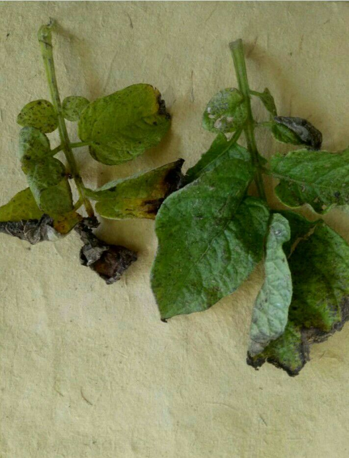
Potato late blight
Symptoms:
Cucumber angular leaf spot primarily affects leaves, but also fruits and stems. On leaves, infection initially manifests as pale green, water-soaked spots, gradually turning yellowish-brown, light brown, pale yellow, grayish-white, and finally white. Due to the restriction of leaf veins, the lesions on the underside of the leaf are polygonal. In humid conditions, a milky-white bacterial ooze appears on the
lesions on the underside of the leaf; when dry, it appears as a thin white film or white powder, brittle and easily perforated. On stems, petioles, and tendrils, infection results in small, water-soaked spots that expand longitudinally along the stem grooves into short strips. Bacterial ooze may also appear in high humidity. In severe cases, the affected area cracks longitudinally, becoming water-soaked and rotten, turning brown, and leaving white marks on the surface after drying.
On fruits, lesions initially appear as small, round, water-soaked spots, expanding into irregular or contiguous lesions. These lesions ulcerate, oozing white bacterial ooze, often accompanied by soft rot fungus infection, resulting in a yellowish-brown, water-soaked rot.
Prevention and control measures:
Crop rotation should be implemented to prevent continuous cropping, especially with solanaceous crops, or near-planting. A rotation of at least 3 years with cruciferous vegetables should be practiced, and planting adjacent to potatoes should be avoided.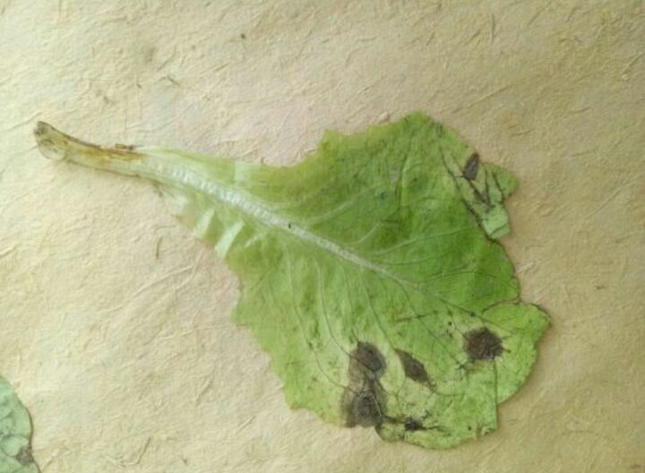
Lettuce sclerotinia disease
Symptoms
This disease primarily affects the base of the stem. Initially, the affected area appears as a yellowish-brown, water-soaked lesion, gradually spreading to the entire stem, causing it to rot. Alternatively, the disease can progress upwards along the leaf margins, leading to rot of the leaf margins and leaves, eventually causing the plant to wilt and die. In protected environments with high humidity, dense, fluffy mycelial masses develop on the affected area, later transforming into black, rat-dung-like sclerotia.
Prevention and control measures
(1) Cultivation and disease prevention. After harvest, thoroughly clean up diseased and fallen leaves, and plow to a depth of 50-60 cm to bury the pathogens deep in the soil, preventing them from germinating or the apothecia from emerging. You can also cover the soil with a mulch film that blocks ultraviolet light to prevent the sclerotia from germinating or to prevent the ascospores from drifting away, thus reducing the primary source of infection.
(2) Soil treatment. After the spring crop is finished, clean up the diseased and fallen leaves, apply 200-300 kg of quicklime and 400-500 kg of chopped rice straw or wheat straw per 667 square meters, then turn the soil, make ridges, water it, and finally cover it with plastic film and close the greenhouse for 7-15 days to keep the soil temperature above 60℃ for a long time to kill harmful pathogens.
(3) Chemical control. Before transplanting, spray the seedbed with 8000 times dilution of 40% Xinxing EC or 4000 times dilution of 25% Fenoxacin WP. In the early stage of disease, first remove diseased plants and leaves, then spray with 600 times dilution of 65% Mancozeb WP, 600 times dilution of 50% Carbendazim WP, 1200 times dilution of 40% Iprodione WP, 500 times dilution of 40% Iprodione WP, or 800 times dilution of 45% Tebuconazole WP. Focus on spraying the stem base and basal leaves. In areas where conditions permit, it is best to use a dust agent.
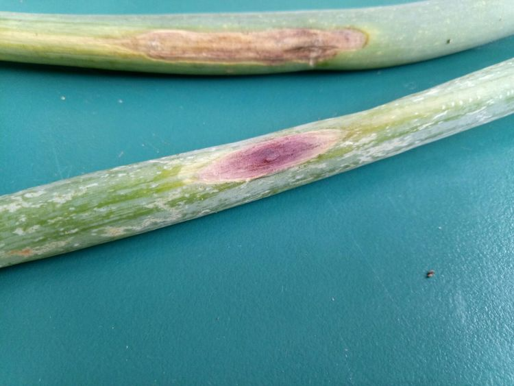
Purple spot disease of scallions
Symptoms:
Primarily affects leaves and flower stalks, occasionally bulbs. In the early stages, lesions are small, gradually enlarging to become oval or spindle-shaped, brown to purple. In humid conditions, a black mold layer develops on the lesions, with concentric rings; the affected area is easily broken. Lesions on scallions and onions are purplish-brown, while those on garlic are yellowish-brown. In high humidity, they are covered with dark brown mold arranged in concentric rings; severely affected plants suffer leaf and flower stalk death. When the bulb and neck are infected after the top has been cut, they exhibit semi-moist rot, turning red or yellow, gradually becoming dark brown or black. The fungus can continue to develop during storage. It belongs to the Deuteromycetes. It affects leaves, branches, and fruits. Leaf lesions are classified into acute and slow-developing types. The acute type forms large lesions on leaves, or causes leaf scorching and significant leaf drop. In the slow-developing type, light brown dots initially appear on the leaves, later merging into irregular large spots with narrow black rings at the edges, a grayish-white or grayish-yellow center, and scattered small black dots. There are two types of lesions on branches: gummosis type and wilt type. Both types exhibit symptoms similar to those of branch and trunk rot, and occur on the root collar, main trunk, and lateral branches. Gummosis type lesions show both soft rot and gum exudation. Wilt type lesions cause the bark to dry out, crack, peel, and eventually fall off severely. Lesions affecting the fruit are round, purplish-brown, and later become distinctly sunken, with scattered small black dots on the surface.
Prevention and control measures:
1. Crop rotation? Implement a crop rotation of 2 years or more with non-onion crops.
2. Chemical control? Suitable fungicides include 70% chlorothalonil wettable powder (600x dilution), 43% cymoxanil suspension (3000-4000x dilution), 70% mancozeb (500x dilution), 75% chlorothalonil (600x dilution), 64% cymoxanil (500x dilution), 58% mancozeb (500x dilution), 70% mancozeb wettable powder (500x dilution), and 64% cymoxanil wettable powder (500x dilution).
3. Strengthen fertilization to enhance tree vigor; avoid excessive humidity inside the greenhouse; and remove fallen leaves and rotten fruit to reduce pathogen sources. Spray with 50% thiophanate-methyl at a dilution of 500-600 times; or 65% zineb at a dilution of 500-600 times; or 80% mancozeb at a dilution of 600 times. From fruit harvest until flowering, 0.3%-0.4% Bordeaux mixture can be used for control.
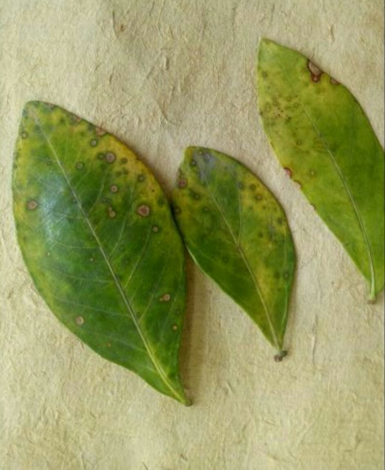
Gardenia anthrax
Symptoms
Anthracnose in gardenias primarily affects the leaves. The disease usually starts on the lower leaves, forming small, round, brown spots that gradually turn yellow and fall off. It can also occur at the leaf tips and edges, forming irregular brown spots that gradually turn brown, harden, and fall off. Severely affected plants may experience significant leaf drop and even die back of branches. Under humid conditions, small black dots appear on both sides of the leaves, with more dots on the underside (the upper surface forms conidiophores, the lower surface forms ascothecia).
Prevention and control measures
1. Promptly remove diseased fallen leaves and destroy them in a centralized manner to reduce sources of infection.
2. Chemical control. Spray with 1% Bordeaux mixture before the onset of disease to protect the plants from infection; during the disease period, spray with 75% chlorothalonil wettable powder at a dilution of 500-800 times.
3. In the early stages of the disease, the root zone soil can be irrigated with a 30-50 times dilution of ferrous sulfate (black alum), or "alum fertilizer water" (2.5 kg of ferrous sulfate, 5 kg of soybean cake, and 15 kg of pig manure mixed and fermented for 10-15 days before application) can be applied alternately with water. Alternatively, a 0.1%-0.2% ferrous sulfate solution can be sprayed on the leaves.
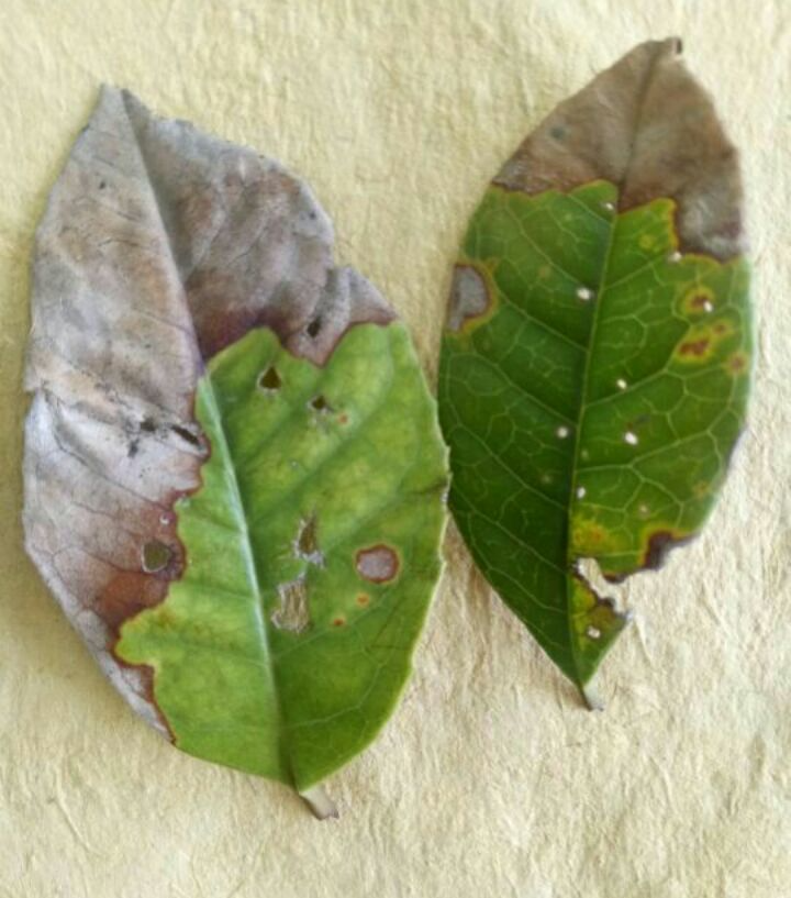
Tea tree leaf blight
Symptoms
Tea leaf blight is a common and serious leaf disease of tea trees. Affected plants grow poorly, with weak buds and premature aging, which seriously affects yield.
Pathogens:
*Colletotrichum camelliae* is a fungus belonging to the subphylum Deuteromycetes, order Colletotrichumales, genus *Colletotrichum*, and species *Colletotrichum camelliae*. The small black dots seen on the lesions are conidiomata, round, 150-330 micrometers in diameter. The conidiophores are short, linear, solitary, without other features, unicellular, and germinate into two cells.
Prevention and control measures
I. Strengthen management and cultivate robust tea plants. Timely cultivation and weeding to loosen the soil; ensure proper drainage in the tea garden or implement drought and cold resistance measures to create an environment conducive to tea tree growth, promote robust tea trees, and enhance disease resistance.
2. Clean up the tea garden to reduce the source of pathogens. For severely diseased tea gardens, diseased leaves should be removed during winter and spring, and dead branches and fallen leaves should be cleared away, or the tea garden should be plowed in winter to bury the diseased leaves deep in the topsoil to block the source of the disease. During the disease outbreak, remove diseased leaves to prevent the spread of the disease.
III. Timely spraying to control the spread of disease. Since this disease is related to leafhoppers, mites, black rot, and red rust, its control should be combined with the control of these pests and diseases. The following pesticides can be used: 0.6% Bordeaux mixture (half-strength lime, harvesting can begin 20 days after spraying), 70% thiophanate-methyl wettable powder at a dilution of 1000-1500 times, agricultural antibiotic actinomycin at 30-50 mg/kg (harvesting can begin 5 days after spraying), antibiotic fenvalerate at 1 mg/kg (harvesting can begin 7 days after spraying), and 80% zineb wettable powder at a dilution of 600-800 times (harvesting can begin 15 days after spraying). The dosage is 60-75 kg per acre, spraying once every 10-15 days, for a total of two sprays. These pesticides can also be sprayed on unharvested tea gardens or nurseries. Depending on the actual situation, the time can be shortened and the number of sprays increased to achieve better disease prevention.
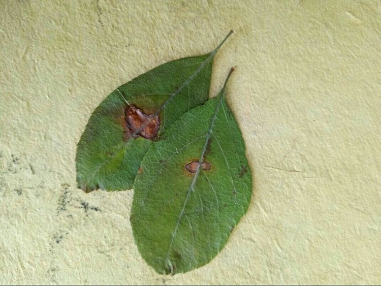
Brown spot disease of Malus halliana
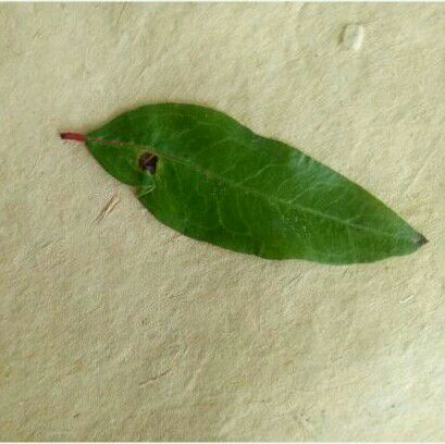
In southern regions, brown spot disease is one of the most common diseases affecting weeping crabapple and a major cause of premature leaf drop. This disease can affect weeping crabapple throughout its entire growth cycle.
In the early stages of the disease, white patches appear on the upper surface of the leaves. These patches then expand, turning brown after about 20 days, and the leaves gradually wither and fall off. Without treatment, the crabapple may lose all its leaves by July and may even bloom a second time. With timely treatment, the leaves can be preserved until the first frost. Therefore, prevention and control of this disease are necessary throughout the entire growth cycle of the crabapple.
Prevention and control methods: First, the planting density should be appropriate. Do not plant too densely, and do not plant in sheltered and shady areas, otherwise the disease will be more severe.
Pomegranate anthrax
Symptoms:
On leaves, semi-circular, nearly circular to elliptical dark brown spots appear mostly at the leaf tip or margin. The edges are dark brown and slightly raised, with a slightly sunken center and faint concentric rings. The spots merge to form patches, causing the leaves to dry out and easily fall off. On shoots, short, slightly sunken dark brown spots initially appear, expanding around the stem and causing the shoots to die. On fruit, irregular dark brown spots appear on the surface, with a slightly sunken center. The spots merge to form patches. Young fruit often dries out and falls off. In nearly mature fruit, as the disease progresses deeper, the flesh also turns brown and rots. In severe cases, the fruit partially or mostly softens and rots, reducing or completely eliminating its commercial value and rendering it inedible. When the weather is humid, small black or vermilion spots appear in a near-ring-shaped arrangement on the affected parts of the leaves, branches, and fruits. These are symptoms of the disease (sporulation discs and conidia of the pathogen).
Control measures:
1. Maintain good field hygiene, especially by thoroughly cleaning the orchard in winter and spring. Collect dead branches, diseased branches, fallen leaves, and diseased fruits and burn them. On this basis, spray the ground and the trees with pesticides once for prevention. This work is even more important for orchards that are prone to the disease.
pear rust
Symptoms
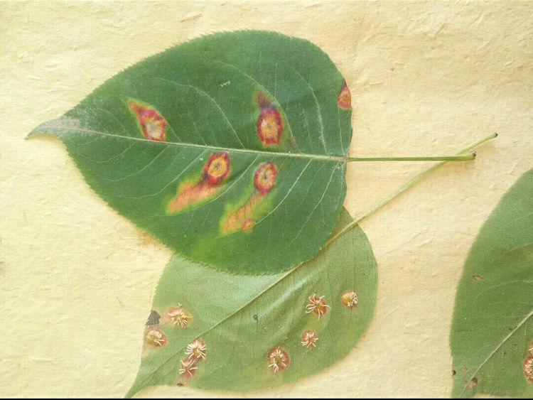
It mainly affects the tender green parts of pear trees, such as young leaves, petioles, young fruits, and new shoots. The key points for diagnosing pear rust can be summarized as: "The affected area is orange-yellow, thickened and swollen, the initial red spots gradually turn black, and later, fine and long yellow hairs grow."
Young leaves: After infection, small, round lesions appear on the upper surface of the leaves. The center is orange-yellow and glossy, while the edges are pale yellow, surrounded by a yellow halo. As the lesions enlarge, honey-yellow, slightly raised dots (fungal cysticerci) appear in the center. When moist, pale yellow mucus (sexual spores) oozes from these dots. After the mucus dries, the yellow dots turn black. Subsequently, the lesion tissue thickens, becoming sunken on the upper surface and raised on the lower surface, from which several to a dozen grayish-white or pale yellow thin tubular structures (aerosporia) grow (affinia). These contain a large number of brown aerospores, which, upon maturation, open and disperse from the top of the aerosporia.
Young fruit: In the early stage of infection, the symptoms are similar to those on the leaves. In the later stage, rust spores appear on the diseased parts. When the disease is severe, the fruit becomes deformed and falls off prematurely.
Petioles and fruit stalks: After being affected, the diseased parts turn orange-yellow and swell up in a spindle shape. Sex organs and ovule cavities may also grow on the lesions.
Prevention and control measures
1. Do not plant junipers within 5 kilometers of the pear orchard to interrupt the transition to a new host plant.
2. If there are junipers near the pear orchard, spray them with lime sulfur solution at 1-2 Baume degrees or sodium pentachlorophenate at 350 times dilution from late February to early March to kill the overwintering teliospores and basidiospores.
3. From the time pear leaves unfold until late May, spray with Bordeaux mixture at a ratio of 1:3:200-240; or 65% zineb at a dilution of 500. Do not spray during the flowering period to avoid phytotoxicity.
Papaya rust
Symptom editing
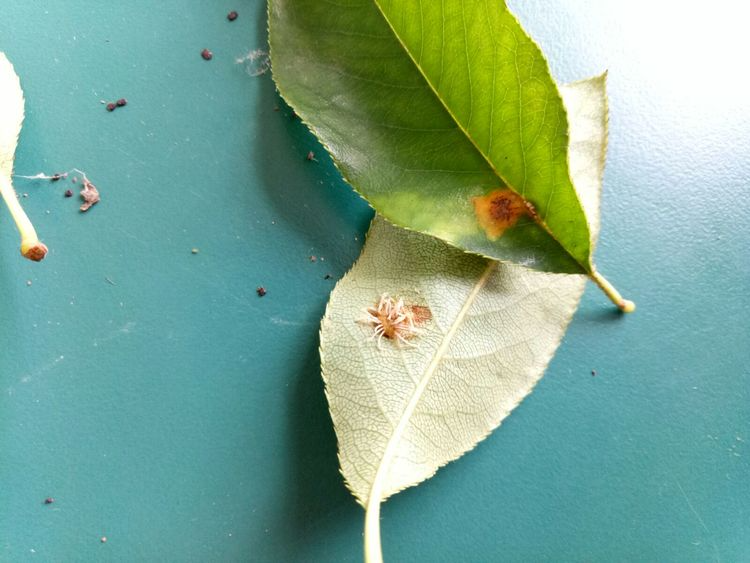
It primarily affects leaves, petioles, tender shoots, and young fruits. Initially, infected leaves develop small, withered yellow spots on the upper surface, which later enlarge into circular patches. The diseased tissue thickens and bulges towards the underside of the leaf, where grayish-brown hair-like structures—the fungal rust spores—grow. These rupture, releasing rust-colored powder. In later stages, the lesions turn black, wither, or fall off. New shoots and young fruits exhibit the same symptoms, becoming deformed or cracked, and are prone to dropping.
pathogen
*Gymnosporangium asiaticum* Miyabe ex Yamada, also known as *Gymnosporangium haraeanum* Syd., is a fungus belonging to the Basidiomycota. This fungus lacks a urediniospore stage. Teliospores are two-celled with a stalk. Sex spores are borne under the epidermis on the upper surface of the leaf, flattened-globose; sex spores are elliptical or fusiform, measuring 5–12 × 2.5–3.5 μm. Aeropspores are borne on the lower surface of the leaf, appearing as hair-like structures, several clustered together. Aeropspores are nearly round, unicellular, yellow or light brown, measuring 19–24 × 18–20 μm.
Transmission routes and conditions for disease development
The fungus overwinters on juniper trees. In March and April of the following year, it produces rice-grain-sized reddish-brown teliospore masses. After rain, these masses swell to form a brown, gelatinous mass. The teliospores on these masses germinate to produce basidiospores, which are then dispersed by wind to quince trees for infection. Later, aeciospores are produced on the lesions, and the released aeciospores are carried by the wind to juniper trees, where they infect and overwinter.
Prevention and control methods
(1) Do not plant juniper within 4km of the papaya orchard, or choose a location far away from juniper to avoid its alternate host and cut off the infection cycle. (2) If necessary, when the papaya sprouts and new leaves appear, spray with 20% triadimefon EC at a dilution of 2000 times or 20% oxychloride EC at a dilution of 200-300 times. In severe cases, spray with 40% fusarium oxychloride EC at a dilution of 9000 times.
Peach tree gummosis
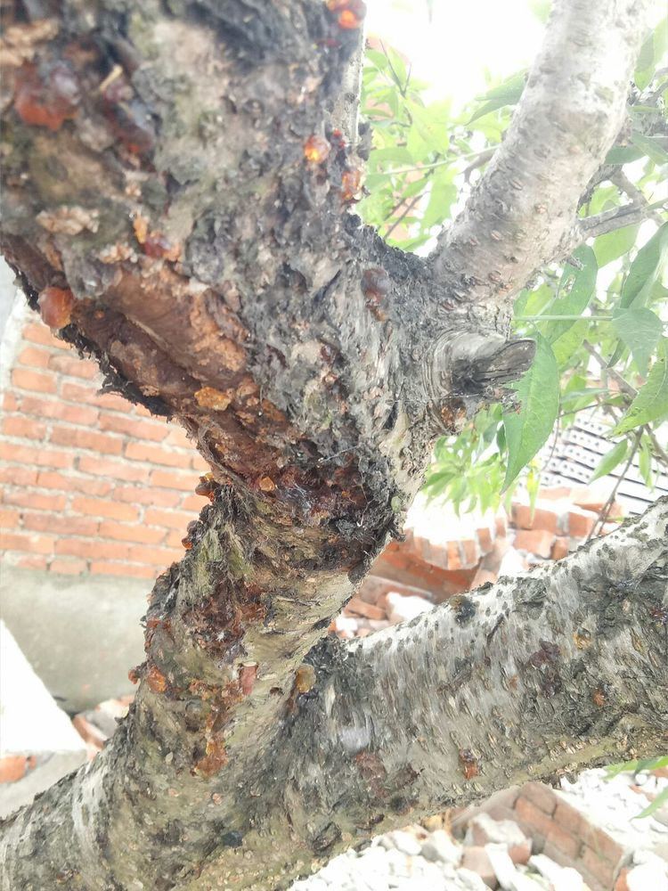
Peach gummosis is divided into two types: infectious and non-infectious. [1]
Non-infectious gummosis of peach trees is a physiological disease, with symptoms similar to those of the former. Its causes include frost damage, pests and diseases, hail, excessive winter pruning, and numerous large mechanical wounds. In addition, excessive fruit production and weakened tree vigor can also induce physiological gummosis.
Prevention and control measures:
① Strengthen fertilizer and water management, and apply 40-80 kg of organic fertilizer per mu. Peach trees are drought-tolerant and suitable for planting in loose soil. Spray with 1000 times diluted tree protectant for sterilization and disinfection as needed. Before the flowers bloom red, apply lime sulfur mixture to the branches for orchard sanitation treatment. Alternatively, use 0.04% brassinolide aqueous solution at 10,000 to 20,000 times dilution to enhance tree vigor and improve disease resistance.
② Master new management techniques for scientific pruning. Pay attention to timely thinning and pruning back during the growing season, and reduce thinning in winter to minimize wounds on branches and trunks. Apply wound-healing and antiseptic film to the pruning wounds promptly to protect them from external bacterial infection and effectively prevent rotting and gumming. Pay attention to thinning flowers and fruits to reduce the fruit load.
③ Prevention: Apply pesticides promptly during the growing season (April-May). Spray every 10-15 days with 1000 times diluted 70% methyl thiophanate wettable powder, or 800 times diluted 72% cymoxanil-mancozeb wettable powder, or 5000 times diluted 43% tebuconazole suspension, or 5000 times diluted 10% difenoconazole water dispersion, combined with 800 times diluted 2% kasugamycin aqueous solution for foliar spraying.
④ For treatment, use 2% kasugamycin aqueous solution diluted 500 times, or add 40% streptomycin wettable powder diluted 10000 times and spray evenly in the field. For severely affected trees, after scraping off the lesions, apply 300 times diluted 2% kasugamycin aqueous solution in combination with streptomycin, which can effectively cure peach tree gumming.
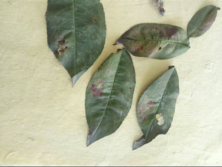
Peach leaf (brown spot) perforation disease
Peach leaf spot disease includes two types: bacterial and fungal. Common symptoms include nearly circular, brown leaf spots that easily fall off, forming perforations, leading to premature leaf drop and weakened tree vigor. Preventative measures include spraying with 1% Bordeaux mixture after spring orchard sanitation. When new shoots emerge, spray with a 1:2:120 lime-based Bordeaux mixture or a 1000-1500 times dilution of 20% quinclorac + 75% chlorothalonil (1:1).
Brown spot perforation: This disease affects leaves, new shoots, and fruits. Circular or nearly circular lesions appear on both sides of the leaves, with purplish or reddish-brown edges and slight ring-like markings, measuring 1–4 mm in size. Later, grayish-brown mold grows on the lesions, the center dries up and falls off, forming a perforation with neat edges, often surrounded by a ring of necrotic tissue. On new shoots and fruits, brown, sunken lesions with reddish-brown edges develop, covered with gray mold.
The pathogen is *Pseudocercospora circumscissa* (Sacc.) Liu & Guo, belonging to the genus *Pseudocercospora* in the subphylum Deuteromycetes. A common synonym is *Cercospora circumscissa* Sacc. Conidiophores are clustered, unbranched, straight or slightly curved, olive-colored, with 0-2 septa, measuring 6.5-35.0 μm × 2.5-4.0 μm. Conidia are slender, whip-like or obclavate, straight or slightly curved, pale bluish-yellow, with 3-9 septa, measuring 25.0-80.0 μm × 2.0-4.0 μm.
Symptoms
Leaves: The lesions are round or nearly round, brown, with reddish-brown edges and slight rings. When moist, grayish-brown mold grows on both sides. Eventually, the diseased parts dry up, fall off, and perforate, with neat edges of the perforations.
Branches and fruits: The lesions are similar to those on leaves, and mold also grows when it is humid.
Disease pattern
Brown spot disease overwinters as mycelium in diseased tissue or latent in diseased shoot tissue.
As temperatures rise the following spring, rain will cause conidia to grow and spread through wind and rain, infecting the body.
Brown spot and shot-hole disease: The pathogen overwinters as mycelium in diseased leaf or twig tissue. The following spring, as temperatures rise and rainfall occurs, conidia are produced and spread by wind and rain, infecting leaves, twigs, and fruits. Subsequent infections are caused by conidia produced in diseased areas. The pathogen develops at temperatures between 7 and 37°C, with an optimum temperature of 5 to 28°C. Low temperatures and abundant rainfall favor the occurrence and spread of the disease.
Prevention and control measures
Strengthen orchard cultivation and management to enhance tree vigor. Apply fertilizer rationally, increase the application of organic fertilizer, and avoid excessive nitrogen fertilizer. For peach orchards with high groundwater levels or heavy clay soil, improve the soil and ensure timely drainage. Perform proper shaping and pruning, combining with winter pruning, promptly remove diseased branches and thoroughly clean up diseased leaves, and burn or bury them deeply. Before peach trees sprout in early spring, spray with lime sulfur mixture or 45% crystalline lime sulfur mixture, 1:1:100 Bordeaux mixture, 30% Green Power suspension, or 80% sodium pentachlorophenate (spraying is best done on sunny, windless days). After leaf expansion, spray with fungicides such as 65% zineb, 50% carbendazim, 70% thiophanate-methyl, 40% chlorothalonil, zinc sulfate lime solution (0.5 kg zinc sulfate, 2 kg lime, 100-125 kg water), 72% agricultural streptomycin, and streptomycin sulfate. During the rainy season of the growing season, spray with mancozeb, captan, or zineb.
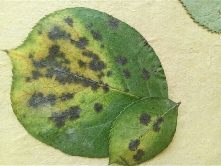
Black spot disease of roses
Rose leaves, young shoots, and flower stalks can all be affected. Initially, leaf spots appear as small purplish-brown to brown dots, later expanding into round spots 1.5–13 mm in diameter, black or dark brown with fibrous edges, although some varieties may have smooth, even edges. A yellow halo often surrounds the spots. Under a magnifying glass, small black pustular granules are visible on the affected area. Several spots often merge, and the surrounding leaves turn yellow over a large area, making the spots appear as "islands" with green edges. Diseased leaves easily fall off, but some rose varieties do not. Purple to black streaks appear on young shoots and flower stalks, slightly sunken. In severe cases, the entire lower and middle leaves of the plant fall off, leaving only a few new leaves at the top. Disease Characteristics: Rose black spot is a fungal disease. It overwinters on diseased plant debris and spreads via rainwater or sprinkler splashes; insects can also transmit it. The optimal temperature for disease development is around 26 degrees Celsius. During rainy seasons, host plants are more severely affected. Newly transplanted plants, those with damaged root systems, and those with weakened growth are more susceptible. In the Beijing area, the disease appears in mid-May, with peak incidence from July to September. In the Yangtze River basin, two peaks occur: May-June and August-September. Control methods include field management : 1. Maintain appropriate planting density and pot density to ensure good ventilation and sufficient sunlight, at least 6 hours per day. 2. Cultivate disease-resistant varieties and use resistant rootstocks, eliminating susceptible varieties with poor ornamental value. 3. In the early stages of disease, promptly remove diseased leaves to reduce the source of reinfection. Avoid touching the leaves of diseased plants to prevent human-mediated spread. 4. Implement preventative measures before disease onset. Prune diseased plants heavily in winter, removing pathogens from stems and branches. Treat fallen leaves and soil in rose planting areas. 5. Reduce sources of infection. In autumn, thoroughly remove dead branches and fallen leaves, and combine this with winter pruning to remove diseased branches. Promptly remove dead leaves and residual branches, and burn them to reduce sources of infection. 6. Improve environmental conditions to control disease occurrence. Irrigation is best done using drip irrigation, furrow irrigation, or watering along the edge of the pot; avoid sprinkler irrigation. Irrigation is best done on sunny mornings to keep the leaves dry. 7. Plant disease-resistant varieties, following normal planting and maintenance procedures. Planting should not be too dense. Water and fertilizer management should be reasonable and scientific, with ample application of phosphorus and potassium fertilizers to promote robust plant growth and enhance disease resistance. Traditional Chinese Medicine Treatment and Prevention: Dilute 30ml of "Su Jing" (速净) with 15kg of water, apply once every 7 days. Key prevention period: Begin spraying when new leaves are just unfolding. Treatment: Dilute 50ml of "Su Jing" + 15-20ml of "Garlic Oil" with 15kg of water, apply once every 3-5 days, for two consecutive applications. After disease control, switch to a prevention plan. Foliar fertilizer application: To improve the photosynthetic rate of leaves and enhance plant immunity, foliar fertilizer - Wofengsu can be sprayed regularly.Black spot disease of roses is a very serious disease. The pathogen damages the leaves, causing a large number of leaves to fall off, resulting in poor plant growth.
Symptoms: Rose leaves, young shoots, and flower stalks can all be affected. Initially, leaf spots appear as small purplish-brown to brown dots, later expanding into round spots 1.5–13 mm in diameter, black or dark brown with fibrous edges, although some varieties may have smooth, even edges. A yellow halo often surrounds the lesions. Under a magnifying glass, small black pustular granules can be seen on the affected area. Several lesions often merge, and the surrounding leaves turn yellow over a large area, making the lesions appear as "islands" with green edges. Diseased leaves easily fall off, but some rose varieties do not. Purple to black streaks appear on young shoots and flower stalks, slightly sunken. In severe cases, the entire lower and middle leaves of the plant fall off, leaving only a few new leaves at the top.
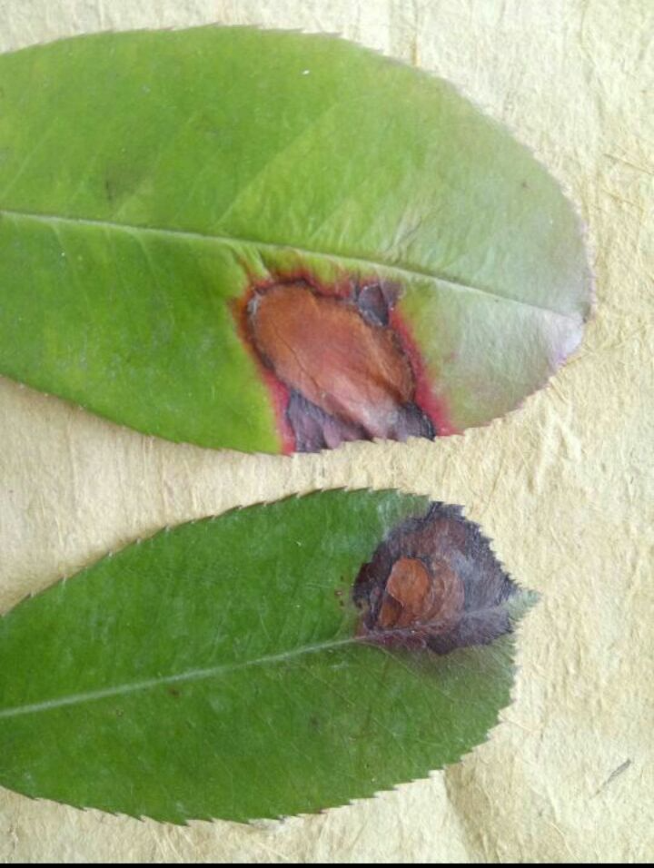
Red photinia anthracnose
Symptoms:
Anthracnose is a major disease of Photinia fraseri during the cutting propagation period. It is more likely to occur in hot, humid, and poorly ventilated environments, and can easily cause Photinia fraseri to only develop roots and not sprout new shoots.
Prevention and control methods:
After the seedling cuttings are completed, pay close attention to the humidity and temperature of the cutting bed, maintain good ventilation, and remove severely affected diseased leaves. In the early stage of disease occurrence, spray with 70% methyl thiophanate wettable powder at a dilution of 500-1000 times or 65% zineb wettable powder at a dilution of 600 times, and spray again after 7-10 days.
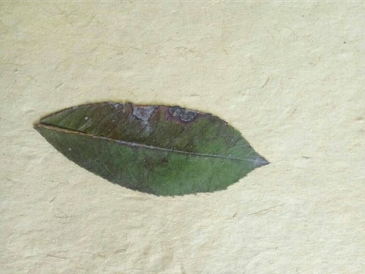
Red Photinia Leaf Spot Disease
Leaf spot disease of Photinia fraseri primarily affects its leaves and stems. When leaves are affected, small brown spots first appear, which gradually enlarge and develop into polygonal lesions. The lesions are reddish-brown on the upper surface of the leaf and yellowish-brown on the underside. In severe cases, the lesions can merge into patches, and the entire plant may even die. This disease is also contagious between plants, posing a serious threat to Photinia fraseri.
Leaf spot disease has many causes, including frequent rainfall and improper planting and management. A large number of pathogens remain in the forests of our region. In densely populated forests, after rain followed by sunshine, a relatively enclosed microenvironment of high temperature and humidity easily forms, further accelerating the invasion and development of leaf spot disease.
Prevention and control of leaf spot disease in red photinia:
1. Improve ventilation in the forest, promptly prune and remove diseased leaves to reduce reinfection.
2. Strengthen management and maintenance, and enhance tree growth and disease resistance through measures such as clearing ditches and draining water, and applying light fertilizer frequently;
3. Select and breed disease-resistant varieties, and avoid continuous planting in the nursery;
4. This disease occurs year-round. Chemical control should focus on prevention. When the leaves first sprout in March and April, spray lime sulfur mixture every 10 days for prevention and protection. In the early stage of the disease, spray with 800 times dilution of 50% carbendazim wettable powder or 500 times dilution of 50% thiophanate-methyl wettable powder. When the disease is severe in spring, spray with fungicide after each rainy season.
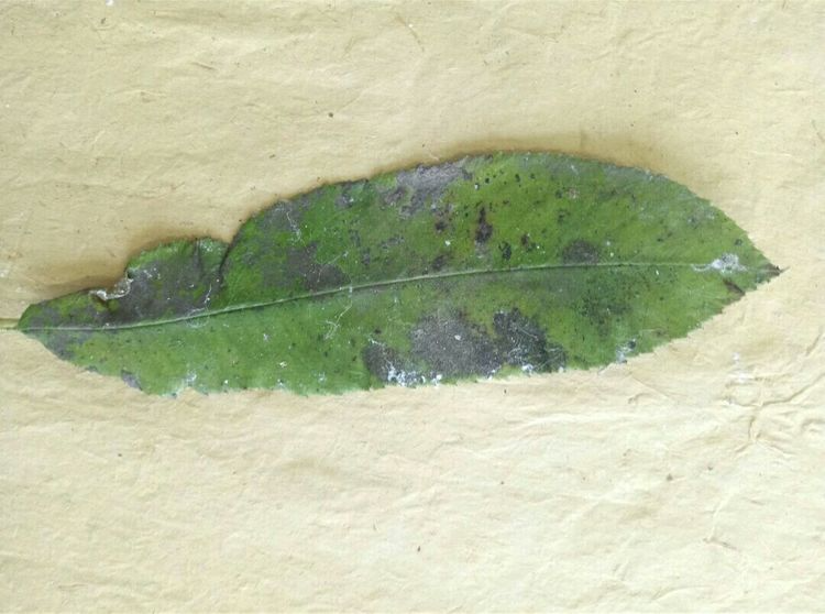
Sooty mold of Photinia serratifolia
Sooty mold, also known as oil spot disease, is a disease caused by insects such as aphids, whiteflies, and scale insects that feed on it by piercing and sucking its prickly flesh. It mainly forms circular black mold spots on the upper surface of leaves and branches, which gradually merge to form patches, covering the entire leaf surface and young shoots with a black mold layer. In severe cases, it causes premature leaf drop.
Control measures: Targeted control measures should be implemented based on the different types of pests, such as aphids, whiteflies, and scale insects.
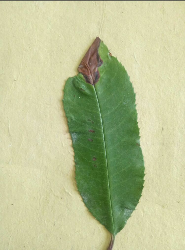
Photinia leaf spot disease
Symptoms:
It primarily damages the leaves. Numerous gray to brown spots of varying sizes appear on the leaves, nearly circular to irregular in shape, ranging from 12-14 mm to 1-3 mm in size. These spots are slightly raised and surrounded by a broad brown line, but do not penetrate the leaf blade. In late winter, small black dots, the conidiophores of the pathogen, appear on the leaf spots. Only small, round, grayish-brown spots are visible on the underside of the leaves, scattered throughout.
Prevention and control methods:
1. In autumn and winter, remove diseased plant debris and burn or bury it deeply to reduce overwintering pathogens.
2. Applying sufficient, well-rotted organic fertilizer to promote vigorous growth can reduce disease incidence.
3. In the early stage of the disease, after removing diseased leaves, spray with Bordeaux mixture (1:1:150), 70% mancozeb (500-800 times dilution), or 50% carbendazim (1000 times dilution). Repeat once every 10 days for 2-3 treatments.
These are some of the symptoms and control methods for leaf spot disease of photinia, compiled by the editor. Have you learned them? Leaf spot disease is quite harmful to photinia, so we need to control it in time to ensure your photinia grows better. If you want to see more knowledge about pest and disease control, remember to follow our pest and disease control knowledge base.
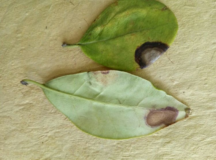
Kanamori Onna-Zai leaf spot disease
Symptoms
It mainly affects the leaves, with lesions that are round to nearly round, 2-6 mm in size, light brown in the center and darker at the edges. Sometimes, a few small brown dots, which are the conidiophores of the pathogen, appear on the lesions.
Transmission routes and conditions for disease development
The pathogen overwinters or hibernates as conidiophores on diseased leaves, and produces conidia the following spring when conditions are suitable for primary and secondary infection. Rainy or humid conditions favor the development of the disease.
Prevention and control methods
Before the onset of disease, spray with 700 times dilution of 75% chlorothalonil wettable powder or 1:1:160 Bordeaux mixture or 400 times dilution of 30% Green Protect suspension.
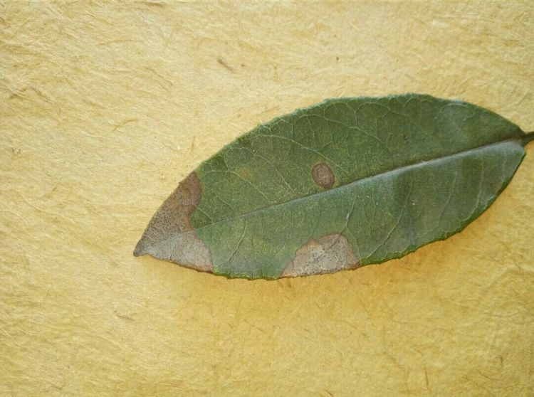
Osmanthus anthracnose
Anthracnose in Osmanthus: The initial lesions on the leaves are small, pale green spots, which enlarge to become round, oval, semi-circular, or irregular in shape, 3–10 mm in size. The center is grayish-brown to grayish-white, and the edge is brown to reddish-brown. In the later stages, small black dots appear scattered, or arranged in concentric rings, which are the conidiophores of the pathogen. When the soil is moist, pinkish mucus is secreted from the small black dots, which is a mixture of pathogen conidia and mucus.
Pathogen: Deuteromycetes, Coelomycetes, Melanosporales, Colletotrichum gloeosporioides
Symptoms of Osmanthus anthracnose: Initially, leaf lesions appear as small, pale green spots. These enlarge to become round, oval, semi-circular, or irregular in shape, 3–10 mm in size, with a grayish-brown to grayish-white center and a brown to reddish-brown edge. Later, scattered small black dots appear, sometimes arranged in concentric rings; these are the conidiophores of the pathogen. In humid conditions, pinkish mucus secretes from these black dots; this is a mixture of fungal conidia and the mucus.
Disease development pattern: The pathogen overwinters as mycelium and conidiophores on diseased leaves and plant debris. Conidia are spread by wind and rain and enter the plant through wounds. The plum rain season in southern China and the rainy season in northern China are the peak periods for the disease. In the Guangzhou area, the disease is generally more severe in late spring and early summer and autumn when there is a lot of rain.
Dissemination method: Spread by wind and rain
Control measures: 1) Remove fallen leaves in winter and disinfect the tree and ground with 1% Bordeaux mixture or lime sulfur solution with a density of 1.002-1.007. 2) Plant osmanthus in fertile, well-drained soil. In orchards where the disease occurs, increase the application of organic and potassium fertilizers. Adjust the density of branches and leaves through pruning and reduce environmental humidity. 3) Spray fungicides at the initial stage of disease. Use 70% copper oxychloride at 300-500 times dilution, 25% carbendazim at 500 times dilution, mancozeb at 500 times dilution, cymoxanil at 500 times dilution, or other fungicides. Various fungicides should be used alternately or in combination.
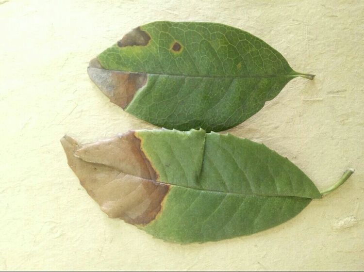
Osmanthus brown spot disease
Osmanthus brown spot disease is a common disease of osmanthus, caused by infection with fungi belonging to the Deuteromycetes. It occurs in both ground-planted and potted osmanthus plants. Mild cases affect the ornamental value, while severe cases can lead to plant death.Morphology and biological characteristics of the pathogen: The stroma is nearly circular, 12–24 μm in diameter, and brown. Conidiophores are unbranched, 0–1-septate, without geniculate nodes, pale olive-brown, nearly colorless at the apex, (3–3.5) μm × (8–30) μm, usually in bundles of 12–32. Conidia are nearly colorless to pale olive-brown, obclavate, with 1–9 septa, erect to slightly curved, obconical at the base, and slightly blunt at the apex, (2.5–4) μm × (10–62) μm. Affected parts: Leaves. Time of infection: This disease occurs from April to October, with more severe outbreaks during rainy seasons and years, and the disease spreads most rapidly in July and August.
Symptoms: Osmanthus brown spot disease is a common disease of osmanthus, caused by infection with fungi belonging to the Deuteromycetes. It can occur in both ground-planted and potted osmanthus plants. Mild cases affect the ornamental value, while severe cases can lead to plant death. Symptoms mostly appear on the leaves, often starting from the leaf tip and margin and spreading to the entire leaf.
Disease Development Pattern: The pathogen overwinters as mycelial masses on diseased and fallen leaves. The following year, under suitable temperature and humidity, it infects and produces conidia, which then infect the plant and cause disease. The pathogen spreads via air currents and water droplets. This disease occurs from April to October, with severe outbreaks during rainy seasons and years, and the fastest spread occurring in July and August. Older leaves are more susceptible than younger leaves; weakened plants and those transplanted in the same year are more prone to infection. Different Osmanthus varieties exhibit varying resistance to brown spot disease; the Dan Gui variety is more resistant than the Jin Gui and Yin Gui varieties.
Transmission route: via airflow and water droplets.
Control measures: 1) Select disease-free plants as propagation mother plants. 2) Strengthen cultivation management, combine with crown shaping, prune weak and diseased branches, adjust the density of branches and leaves, and enhance tree vigor. 3) When seedlings are removed from the nursery, remove diseased leaves and spray with a 1000-fold dilution of potassium permanganate for disinfection. 4) During the early stage of disease in the growing season, spray the entire tree with a protective agent, such as 300-500 times dilution of 70% carbendazim, 1000-1500 times dilution of azoxystrobin, or 500-600 times dilution of 50% carbendazim.
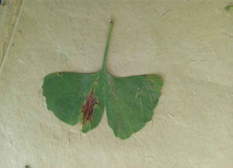
Ginkgo leaf blight
Symptoms of Ginkgo Leaf Blight
In the early stages of the disease, the leaf tips commonly turn yellow. Around June, the yellowed areas gradually turn brown and die, spreading from localized areas to the entire leaf margin, forming brown to reddish-brown leaf margin lesions. Subsequently, the lesions gradually spread towards the base of the leaf until the entire leaf turns brown or grayish-brown, withers, and falls off. From July to August, the boundary between the lesions and healthy tissue is clear, with wavy edges and a darker color. Narrower or wider bright yellow lines can also be seen along the outer edge. From September onwards, the lesions enlarge significantly, and the edges become uneven, with the boundary between diseased and healthy tissue gradually becoming indistinct. In addition, from September to October, irregular discolored spots appear on leaves of shoots sprouting from the base of seedlings or large trees at various locations. These spots are brown in the center, and although they do not enlarge significantly, they often merge with the extending leaf margin spots. During the disease period, various black or grayish-green fuzzy fungal fruiting bodies, i.e., small black dots, can be seen on the lesions. In Jiangsu Province, ginkgo leaf blight occurs around mid-June. After infection, the leaf tips initially show localized browning and necrosis, which soon spreads to the entire leaf tip, appearing as brown or reddish-brown lesions. Subsequently, the lesions continue to extend towards the leaf base, turning dark brown or grayish-brown until the leaf withers and falls off. From June to August, the edges of the lesions are wavy, clearly defined, and sometimes have yellow lines. In September, when the edges of the lesions show water-soaked, seeping expansion, the boundaries between the lesions often become indistinct. During the disease period, various black to grayish-green fuzzy or small black dot-like fungal fruiting bodies can be seen on the lesions.
Prevention and control methods
First, select a site with good conditions for seedling cultivation or planting, avoiding the use of poor, compacted, or waterlogged soil. During cultivation management, attention should also be paid to controlling underground pests and improving cultivation techniques to promote healthy plant growth and enhance disease resistance. Furthermore, ginkgo should not be planted near dawn redwood, and should never be mixed with dawn redwood. In young ginkgo forests, intercropping with broad beans in spring and soybeans in autumn is beneficial to ginkgo growth and can reduce the occurrence of leaf blight. In nurseries with the necessary facilities, spraying with a 400-fold dilution of 40% carbendazim during the disease period can also achieve good control results.
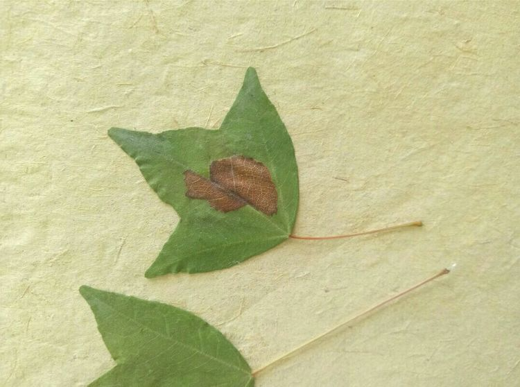
Triangle maple leaf spot disease
The pathogen causing lacquer spot disease in Acer triangularis is *Vladracula annuliformis*. This fungus is distributed throughout Acer triangularis cultivation areas across China.
Symptoms:
In the early stages of the disease, small, chlorotic spots appear on the leaves. The centers of these spots are brown, with purplish-red edges. These spots gradually enlarge into large, round, nearly round, or spindle-shaped lesions. In the later stages, numerous small, raised, black, plaster-like spots appear on the leaf surface, often irregular in shape with rounded edges, eventually causing premature leaf drop. The pathogen overwinters as mycelium and conidia on diseased plant debris. The following spring, apothecia and ascospores are produced and spread by air currents and water droplets starting from the rainy season. Primary infection occurs via ascospores. Black spots appear on diseased leaves starting in mid-to-late August. The initial overwintering inoculum is the most crucial condition for disease development; overcrowding and poor ventilation can contribute to the disease's occurrence.
Prevention and control methods:
1. In autumn and winter, collect diseased branches, leaves and fallen leaves, and burn them promptly to eliminate the source of infection.
2. Starting in May, spray with a 100-200 times diluted Bordeaux mixture every 10-15 days, repeating 2-3 times to prevent disease. (Plant Network)
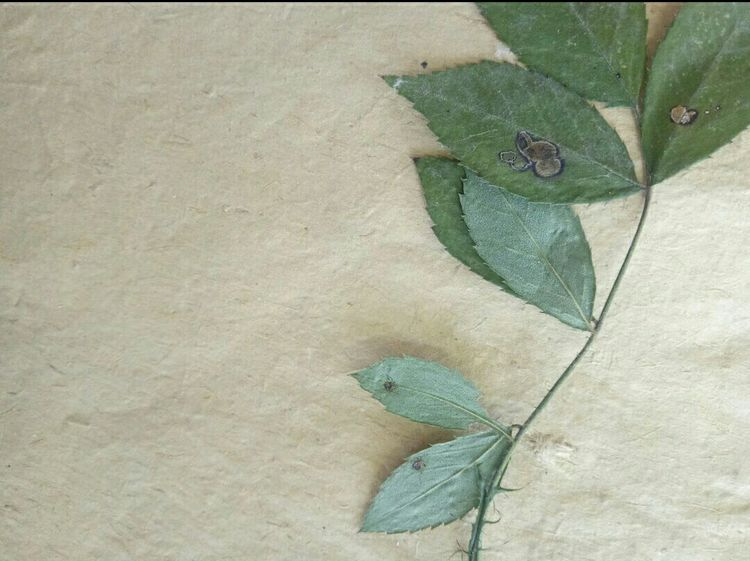
Rose anthracnose
Symptoms
When roses are infected with anthracnose, semi-circular lesions appear on the edges of the leaves, and later, many small black spots appear on these lesions. The disease is more likely to occur when the environment is humid and the plants are too dense.
Anthracnose occurs sporadically and can spread from a single leaf to the entire plant.
Prevention and control methods
In autumn and winter, promptly clean the plants, removing fallen leaves and dead branches. Properly prune the branches to increase light penetration.
When the disease occurs, spray the fungicide promptly. Daconazole, carbendazim, and chlorothalonil are all acceptable. Dilute them with water before spraying. Alternating between several fungicides will yield better results.
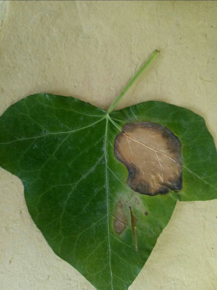
Virginia creeper leaf spot disease
Symptoms
In the early stage of the leaf spot disease of Virginia creeper, small yellow-brown spots appear on the leaves, which later expand into nearly circular spots with a diameter of 3-6 mm. In the later stage, the lesion tissue turns light yellow, but the leaf margin is brown. Small black dots-conidiophores are scattered on the lesions. It can cause the Virginia creeper to wither and fall off prematurely. [2]
Pathogen: Virginia creeper leaf spot disease
The pathogenic fungus of Phyllosticta hedenicola Dur. et. Mont is Phyllosticta hedenicola Dur. et. Mont. The conidia are oval-shaped and grow in the conidiophores, with short gum filaments at one end. [3]
Occurrence pattern
Leaf spot fungus overwinters as mycelium, conidiophores, or ascocarps on diseased parts and becomes the primary source of infection for the following year. It generally occurs in April and peaks from June to August. [4]
Prevention and control methods
1. It can be sprayed with 1% Bordeaux mixture, 50% thiophanate-methyl wettable powder at 800 times dilution, 50% carbendazim wettable powder at 1000 times dilution, or 75% chlorothalonil wettable powder at 800-1000 times dilution.
2. If the spots caused by leaf spot fungus on home-grown Virginia creeper are small and few, you can use a small pin to prick the spots (it's best to sterilize the pin). Then apply a medicated cream (such as Daktarin) to the spots on the Virginia creeper's leaves to control the development of the disease and prevent its spread.
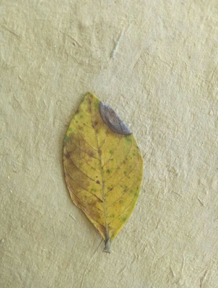
Gardenia chlorosis
Leaf chlorosis first occurs on the tender leaves at the branch tips, starting from the leaf margins and progressing towards the center. The leaf color changes from green to yellow, gradually worsening, with the leaf tissue turning yellow or light yellow, while the veins remain green. Later, the entire leaf turns yellow, then yellowish-white, and finally white, with grayish-brown to brown edges, eventually leading to necrosis and drying. The top leaves are most severely affected, while the lower leaves are normal or nearly normal. In severely affected areas, the plant weakens year by year and eventually dies.Disease Pathogen:
This disease is caused by unsuitable cultivation conditions, such as overly clayey soil, excessive lime, high alkalinity, low-lying and damp conditions, and insufficient iron supply. It is an important physiological disorder.
Occurrence Pattern :
It is more likely to occur in calcareous soil areas.
Control Methods:
1. Horticultural Control: Use well-drained, loose, fertile acidic soil for cultivation. When potting, acidic soil such as mountain soil can be used. Change the potting soil every 1-2 years. Use organic fertilizer, mixing ferrous sulfate and zinc sulfate into the composting process.
2. Chemical control: In the early stages of the disease, irrigate the affected plants with a 2%-3% ferrous sulfate solution, or spray the leaves with a 0.1%-0.2% ferrous sulfate solution, or use an iron compost in the soil (0.2 grams for a 22 cm (6-inch) pot). Chemical treatment for chlorosis should be carried out in the early stages of the disease; otherwise, the effect will be poor.
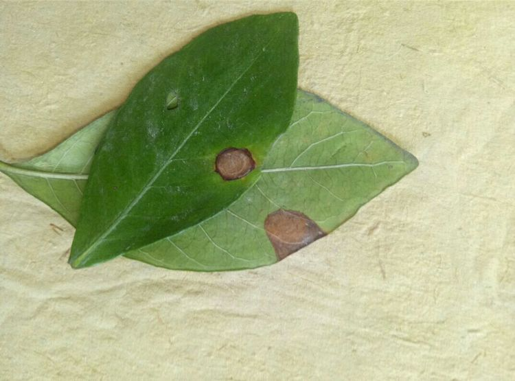
Gardenia leaf spot disease
Leaf spot disease is caused by a fungus and primarily affects the leaves of gardenias. When infected, circular lesions 2-4 cm in diameter appear on the plant, turning yellowish-brown in color, with numerous small black dots appearing along the leaf edges. A humid environment increases the likelihood of this disease in gardenias. Control measures include ensuring good ventilation and light, watering appropriately, and reducing air humidity. If possible, place the plant outdoors. In the early stages of infection, promptly remove diseased leaves to prevent further spread. Separate infected gardenias from other plants to avoid further infection. When the disease occurs, spray with Bordeaux mixture or zineb solution every 7-10 days, for 3-4 consecutive applications to cure the disease.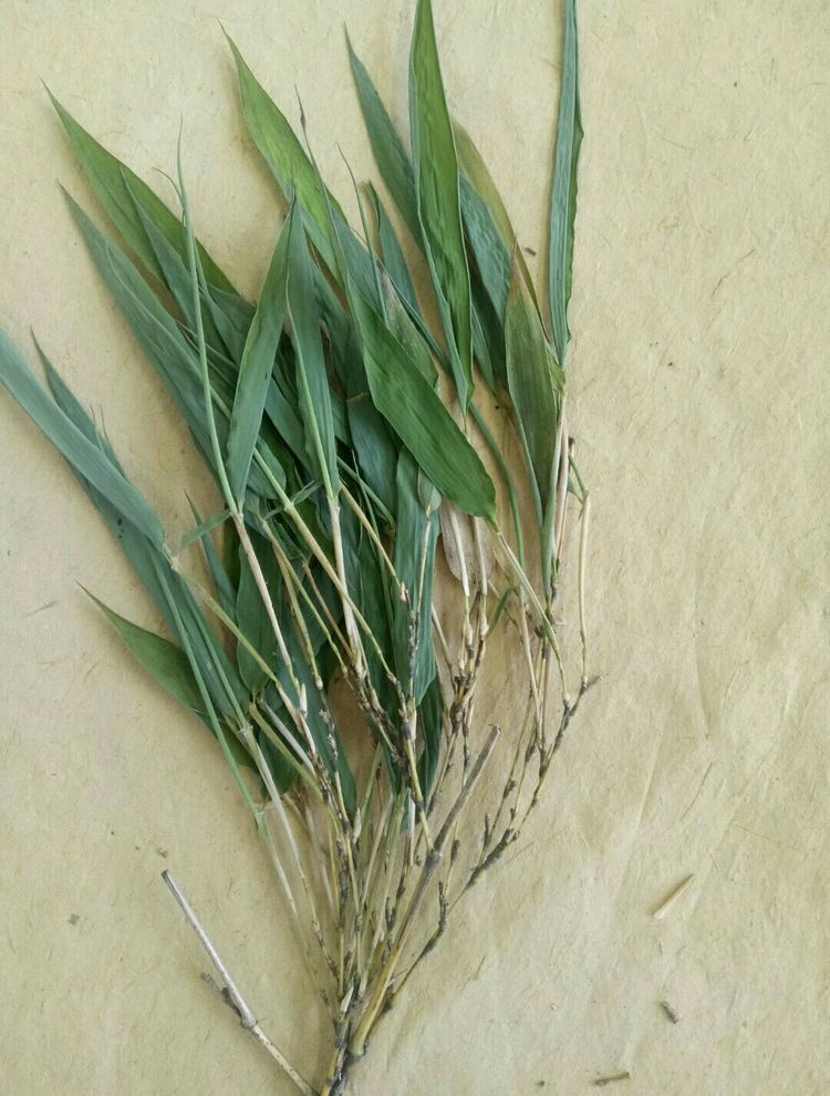
Bamboo bush disease
In the early stages of the disease
, only a few bamboo branches are affected. In spring, these diseased branches continuously extend into thin, multi-segmented vines. Between April and June each year, white, rice-grain-like substances, measuring 5-8 x 3 mm, appear inside the leaf sheaths at the tips of the diseased branches. Sometimes, between September and October, these same white, rice-grain-like substances also appear inside the leaf sheaths at the tips of newly grown diseased branches. The disease initially affects only a few bamboo branches, gradually spreading to all branches over several years.
pathogen
The pathogenic amycota is *Balansiatake (Miyake) Hara*, belonging to the genus *Balansia*, subphylum Ascomycota, order *Pyrophyllales*. The pathogen forms white, nonfunctional granular pseudostruciformes at the tips of diseased branches, formed by the mycelium and host tissue.
Occurrence pattern
It is speculated that the disease may be transmitted through contact. The disease develops from individual bamboo branches to others, spreading from a single point to a patch. Sometimes, short and weak young bamboo shoots grow from mature rhizomes. This disease is more likely to occur in old bamboo forests and poorly managed, weak-growing virgin forests. Bamboo older than four years, or bamboo in areas with strong sunlight, is also susceptible to the disease.
Prevention and control methods
1. Strengthen the nurturing and management of bamboo forests, regularly burn firewood, press soil and apply fertilizer to promote the growth of new bamboo.
2. Cutting off diseased plants as early as possible and repeating this process year after year can yield good results.
3. When establishing new bamboo forests, mother bamboo should not be dug up in the diseased area.
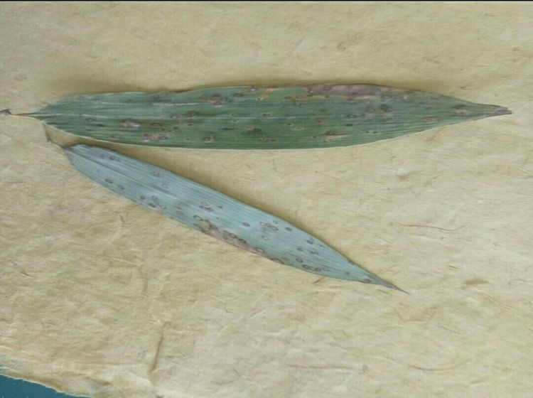
Bamboo black nevus
Distribution and Damage:Black spot disease, also known as leaf spot, is prevalent in bamboo-producing areas. Infected bamboo exhibits stunted growth, with diseased leaves easily withering and falling off, and reduced shoot production.
Symptoms
: In the early stages, small, pale white spots appear on the infected leaf surface, later expanding into round, oval, or spindle-shaped lesions, which gradually turn orange-yellow to reddish. In the later stages, small, shiny, raised black dots, the stromata of the pathogen, appear on the lesions. A distinct orange-yellow discoloration ring surrounds these dots. Lesions can merge to form irregular shapes. Eventually, the diseased leaves partially or completely turn brown and die.
Pathogen: The
pathogen is a variety of *Phyllachora* spp., belonging to the Ascomycota, Rhizobium, and Sphaerocephales.
Occurrence Pattern:
The pathogen overwinters as mycelium or stromata in diseased leaves. The fruiting bodies mature in April and May of the following year, releasing ascospores, which are spread by wind and rain. Infected bamboo begins to develop on leaves near the ground and then gradually spreads upwards.
Prevention and control methods
: ① Reduce sources of infection: In early spring, collect and destroy diseased branches and leaves. ② Strengthen cultivation and management: Loosen the soil and fertilize in a timely manner to promote bamboo growth and enhance disease resistance. ③ Chemical control: At the initial stage of disease, spray with 1:1:100 Bordeaux mixture, or 75% chlorothalonil, or 50% thiophanate-methyl at a dilution of 500-800 times, once every 10-15 days, for 1-2 times.
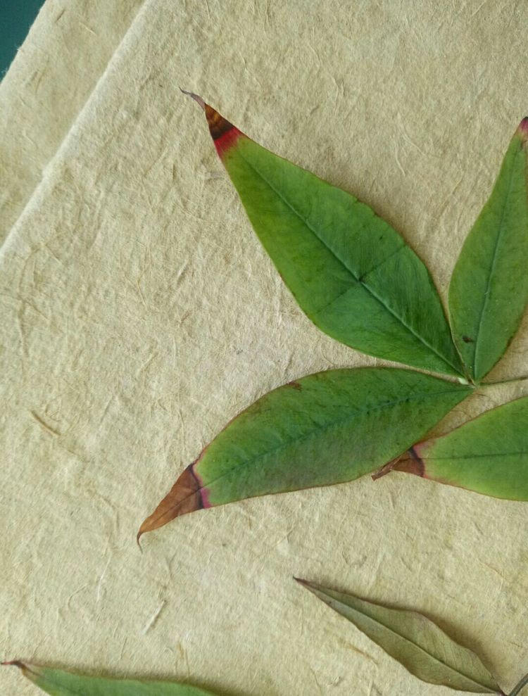
Nandina domestica red spot disease
Symptoms
It usually starts at the leaf tip or edge, initially appearing as small brown spots that gradually enlarge into semi-circular or wedge-shaped lesions, 2-5 mm in diameter, brown to dark brown, and slightly radiating. In later stages, grayish-green to dark green sooty masses, namely conidiophores and conidia, grow on the lesion clusters. Severe disease often causes premature leaf drop.
pathogen
Cercospora nandinae Nagato-mo., a fungus belonging to the class Hyphomycetes and order Hyphomyales.
Occurrence pattern
The fungus overwinters as mycelium or fruiting bodies on diseased leaves, and produces conidia the following spring, which are spread by wind and rain to infect and cause disease.
Prevention and control methods
1. Remove diseased leaves promptly and destroy them or bury them deeply in the soil.
2. In spring, spray with a 400-600 times dilution of 70% zineb wettable powder or a 1000-1500 times dilution of 70% thiophanate-methyl wettable powder for control. Spray once every 10-15 days, for 2-3 consecutive times.
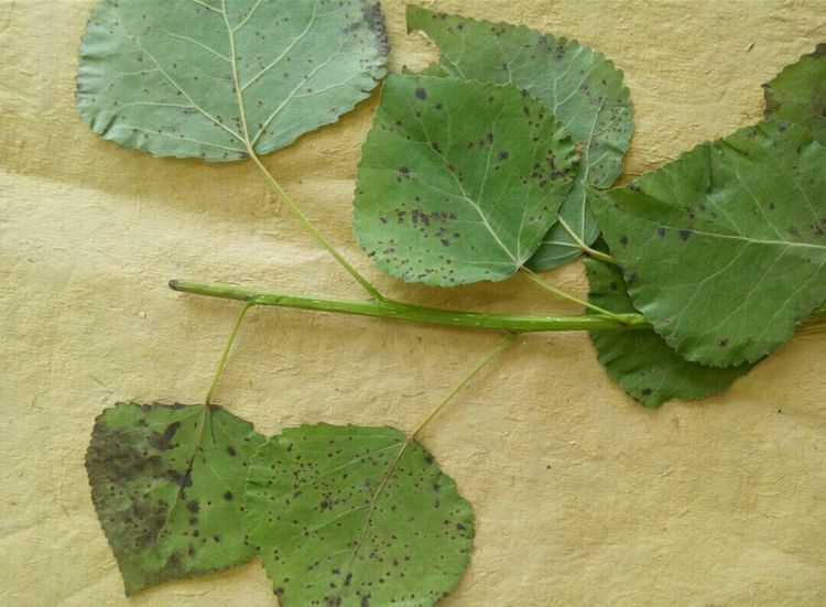
Poplar black spot disease
Distribution and hazards
Poplar black spot, also known as poplar brown spot, is caused by a pathogen that can infect various types of poplar trees, including seedlings, saplings, and mature trees. Severe infection can affect the normal growth of trees and cause economic losses.
Occurrence pattern
The pathogen overwinters as mycelium in lesions on fallen leaves or twigs. The disease begins in early May, peaks in summer and autumn, and persists until leaf fall. It affects poplar leaves, petioles, fruit spikes, and young shoots, forming angular, nearly circular, or irregular dark brown lesions, approximately 1 mm in diameter, sometimes reaching 5 mm. Multiple lesions can merge into large, irregular patches, causing premature leaf drop. The following May and June, newly produced conidia are spread by wind, landing on seedling leaves and entering through stomata. Symptoms appear in 3-4 days, and conidiophores form in 5-6 days, leading to reinfection.
Prevention and control methods
① Strengthen nursery management. Disease-resistant varieties should be selected for seedling cultivation. Pay attention to timely thinning, improve ventilation and light conditions, and ensure proper drainage and other field management practices to reduce the conditions for disease development. Avoid continuous cropping in the nursery or setting up the nursery near susceptible plants. Planned crop rotation for seedling cultivation is recommended. Seeds carrying pathogens need to be chemically treated to prevent disease in seedlings. Dry seeds can be treated with 1000-1500 times dilution of 85% chlorothalonil wettable powder or with methyl thiophanate and carbendazim powder.
② Plant at appropriate density and prune in a timely manner to maintain ventilation and light penetration within the forest. Clean up fallen leaves in the forest promptly to reduce sources of disease.
③ Chemical control: Regularly spray with 600-800 times diluted Guoguang Yintai (80% zineb wettable powder) + Guoguang Sitaling (amino acid chelated multi-trace element foliar fertilizer) for disease prevention and nutritional supplementation, and to improve ornamental value;
In the early stages of the disease, spray with a 500-600 times dilution of 25% imazalil EC (such as Guoguang Bixian) or a 400-600 times dilution of 50% mancozeb WP (such as Guoguang Yingna). Apply 2-3 times consecutively, with an interval of 7-10 days.
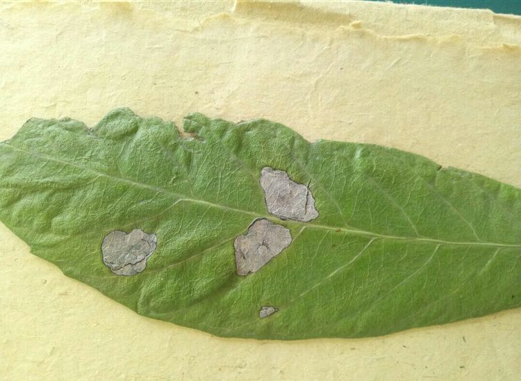
Loquat gray spot disease
Symptoms
This fungus belongs to the Deuteromycetes. It infects leaves, branches, and fruits. Leaf lesions are classified into acute and slow-developing types. The acute type forms large patches of lesions on leaves, or causes leaf scorch and significant leaf drop. In the slow-developing type, light brown round spots initially appear on the leaves, later merging into irregular large spots with narrow black rings around the edges, a grayish-white or grayish-yellow center, and scattered small black dots. Branch lesions are classified into gummosis and wilt types, both with symptoms similar to branch and trunk rot. They occur on the root collar, main trunk, and lateral branches. Gummosis lesions present with soft rot and gum exudation. Wilt lesions cause the bark to dry out, crack, peel, and severely flake off. Lesions affecting fruits are round, purplish-brown, later becoming distinctly sunken, and scattered with small black dots.
Prevention and control methods
Strengthen fertilization to enhance tree vigor; avoid excessive humidity inside the greenhouse; and remove fallen leaves and rotten fruit to reduce disease sources. Spray with 50% thiophanate-methyl at a dilution of 500-600 times; or 65% zineb at a dilution of 500-600 times; or 80% mancozeb at a dilution of 600 times. From fruit harvest until flowering, 0.3%-0.4% Bordeaux mixture can be used for control.
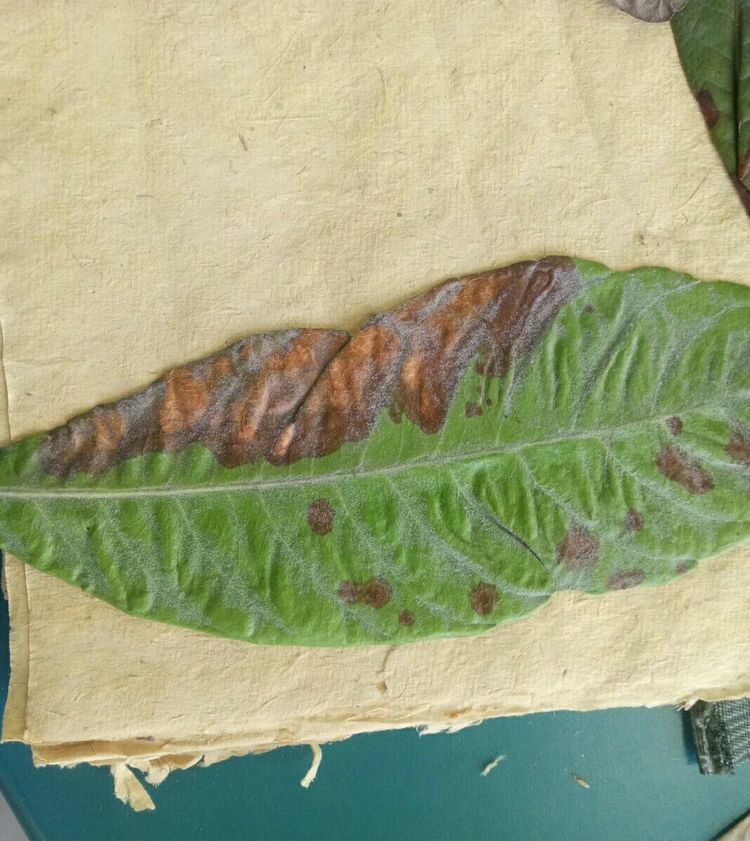
Loquat brown spot disease
Symptoms of loquat brown spot disease:
Only the leaves are affected. Initially, lesions appear on the leaf surface as small, pale yellow spots, which then develop into small brown spots, brown spots, and eventually reddish-brown spots. The lesions are 1-3 mm in size, nearly circular, angular, or irregular in shape. Later, the disease spreads across both sides of the leaf, with the center of the lesions on the leaf surface turning grayish-brown and developing black mold-like granules, which are the conidia and stromata of the pathogen. The most distinctive feature of this lesion is the yellow halo around its outer edge, making it particularly noticeable on the green leaf surface.
[Transmission routes and conditions for the development of loquat brown spot disease]:
The pathogen overwinters as mycelium on diseased leaves, producing conidia the following spring for primary infection. In warmer regions, the fungus can produce conidia year-round, spreading via wind and rain, resulting in multiple reinfections. Poorly fertile, poorly drained, or moisture-stagnant soil exacerbates the disease. Resistance varies among different varieties.
[Prevention and control methods for loquat brown spot disease]:
(1) Plant disease-resistant varieties.
(2) Remove diseased fallen leaves in late autumn and early winter to reduce sources of infection.
(3) Spray 25% benomyl, cyclohexane zinc emulsifiable concentrate at 800 times dilution, or 50% thiophanate-methyl·sulfur suspension at 800 times dilution, or 50% cymoxanil wettable powder at 900 times dilution in the early stage of the disease.
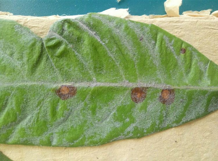
Loquat spot disease
Symptoms of loquat spot disease:
It damages leaves. Initially, the lesions appear as small reddish-brown spots, which gradually enlarge into nearly circular shapes with a grayish-yellow center and reddish-brown outer edges. Most lesions heal into irregular shapes. Later, fine black dots, or conidiophores, appear on the lesions, sometimes arranged in concentric rings. Severe infection can cause premature leaf drop.
[Loquat spot disease infection cycle]:
The pathogen overwinters as conidiophores and mycelium on diseased leaves. In March and April of the following year, after the conidiophores absorb water, conidia are released from the openings and spread by wind and rain, invading and damaging the host.
【Pathogenesis of Loquat Spot Disease】:
It can cause multiple infections within a year, with the most severe cases occurring during the rainy season.
[Prevention and control methods for loquat spot disease]:
(1) Agricultural control: Select disease-resistant varieties. In winter, all fallen diseased leaves and fruits should be completely removed and burned. Strengthen orchard management. During the rainy season, ensure proper drainage to prevent waterlogging, increase soil oxygen supply, and improve disease resistance. In spring, deep plow and expand planting holes to improve soil quality, apply fertilizer appropriately, and mound soil to promote vigorous plant growth. After harvesting, prune weak, dead, diseased, insect-infested, dense, crossing, and overly vigorous branches. After a certain number of years of growth, fruiting branches will have no leaves at the base and will be too long, which will affect fruit development and quality. After harvesting the fruit, prune the branches 10 cm from the base. This will reduce canopy closure and enhance light and ventilation in the canopy.
(2) Chemical control: From the emergence of new shoots to the growth period, use 70% thiophanate-methyl at 800-1000 times dilution, or 50% carbendazim at 800-1000 times dilution, or 0.5%-0.6% Bordeaux mixture, or 77% copper oxychloride at 600-800 times dilution, 12% copper oxychloride at 500-600 times dilution, or 65% zineb at 500-600 times dilution, etc. If 2-3 of these are used alternately, the effect will be better.
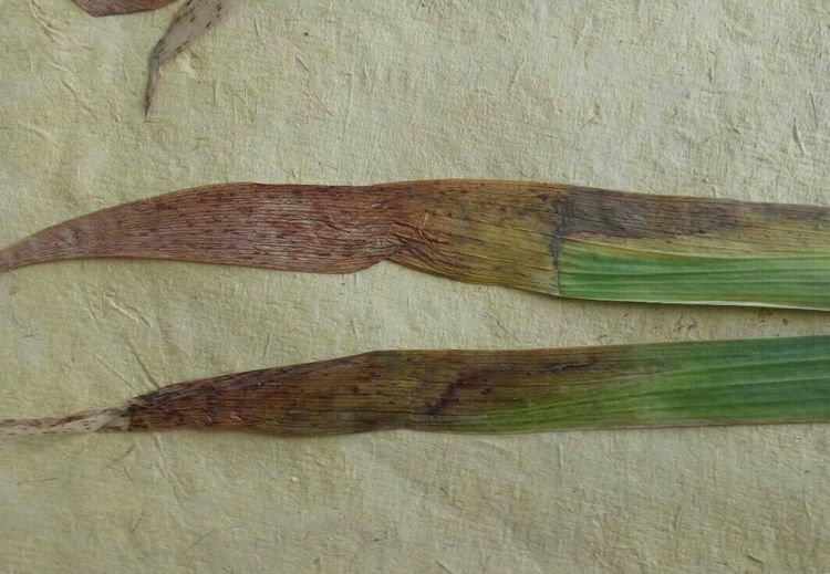
Iris leaf blight
Distribution and damage of iris leaf blight
Iris leaf blight is a common disease that is highly contagious and has a significant impact on iris growth.
Symptoms
It usually starts from the leaf tip. In the early stage of the disease, there are grayish-brown spots or patches of dryness at the leaf tip. Later, it gradually spreads to the base of the leaf, eventually causing the entire leaf to become scorched and black granular substances to appear.
pathogen
The pathogen is a short rod-shaped bacterium.
Prevention and control methods
Strengthen quarantine measures and do not introduce diseased plants. Disinfect the soil and plants before planting. Strengthen daily maintenance and prevent waterlogging in the planting area. Remove diseased leaves promptly at the initial stage of disease to reduce the source of infection. Remove leaves from the ground in winter and burn them. If disease occurs, alternately spray with 800 times dilution of 75% chlorothalonil wettable powder and 1500 times dilution of 70% thiophanate-methyl wettable powder, for 4 to 5 consecutive applications, with an interval of 8 to 10 days between each application.
Symptom identification: The tips of affected liriope leaves begin to turn yellow and brown, gradually spreading towards the leaf base, with a darker color at the boundary between diseased and healthy tissue; sometimes water-soaked spots of different colors appear on the leaves. In the later stages of the disease, the entire leaf turns yellow and dies, severely affecting yield and quality.
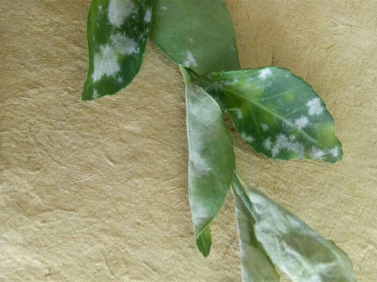
Buxus powdery mildew
Buxus macrocarpa is most susceptible to powdery mildew on its young leaves and new shoots. The most obvious symptom is that the upper or lower surfaces of the leaves and the surface of the young shoots are covered with a white powdery substance, which gradually turns into a whitish-gray felt-like substance in later stages. In severe cases, the leaves curl, the branches twist and deform, and the branches may even die.
Powdery mildew primarily affects the leaves of Buxus macrocarpa, with lesions mainly distributed on the leaf surface, producing white powdery mildew. In severe cases, it can also occur on the underside of leaves and new shoots, leading to wrinkled and deformed leaves, twisted and withered new shoots, and ultimately, leaf drop and shoot dieback. In the early stages, numerous small, white, circular spots appear scattered on the leaves. As these spots enlarge and merge, they become irregular large spots, sometimes covering the entire leaf surface. If the white powdery layer on the surface of the lesions is wiped away, yellow circular spots appear at the affected area. The pathogen overwinters as mycelium (a gray, membranous layer) within the affected tissues of Buxus macrocarpa or between bud scales. The following spring, during the leaf unfolding and growing season, a large number of conidia are produced and spread by air currents, becoming the primary source of infection. The pathogen parasitizes the surface of the host's branches and leaves, producing haustoria that penetrate deep into the epidermal cells to absorb nutrients. Each spring, summer, and autumn, a large number of spores are produced, repeatedly infecting leaves and new shoots. High summer temperatures are unfavorable for disease development. High temperature is a major factor in the occurrence of the disease.
Prevention and control measures:
1. Improve cultivation conditions. The planting site should be high, dry, with sufficient sunlight, free from waterlogging, and with deep and fertile soil.
2. Strengthen fertilizer and water management, increase the application of phosphorus and potassium fertilizers, and apply an appropriate amount of nitrogen fertilizer; pay attention to drought resistance and drainage to ensure the seedlings grow strong and improve their disease resistance.
3. Combine pruning with removing diseased branches and leaves. Thoroughly clean the orchard in winter, sweeping up and burning diseased branches and leaves; promptly prune and burn any diseased leaves and branches discovered during the growing season to effectively reduce the initial and secondary sources of infection.
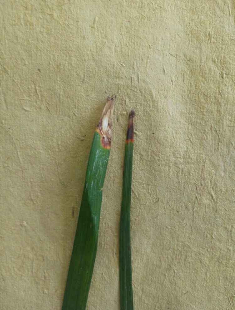
Ophiopogon japonicus black spot disease
Pathogen: *Alternaria* sp. belongs to the subphylum Deuteromycetes, class Hyphomycetes, order Alternariales, family Meliomycetes, and genus *Alternaria*. Conidiophores are solitary or in clusters of 2-30, dark brown, paler at the apex, with slightly larger basal cells, unbranched, straight or slightly curved, without geniculate nodes, and have 2-9 septa, measuring 15-90 μm × 45 μm. Conidia are pale or in chains of 2-3, brown, obclavate; the beak is short to slightly long, slightly paler, and unbranched. The spore body has 2-7 septa and 1-6 longitudinal septa, constricted at the septa, measuring 23-52 μm × 9-12 μm. The beak has 0-2 septa, measuring 5-20 μm × 3-4 μm.
IV. Disease Development Pattern: The pathogen overwinters on seedlings and begins to cause disease in mid-April of the following year. The occurrence and development of the disease are greatly affected by rainfall, with severe outbreaks occurring during the rainy season. In the field, distinct central diseased plants can be observed, which rapidly spread to the surrounding areas. Under suitable temperature and humidity conditions, the disease quickly becomes widespread, causing large areas to wither and die.
V. Prevention and Control Measures: (1) Select healthy, disease-free plants with bright green leaves as seedlings. (2) Remove the central diseased plants in time and replace them with healthy seedlings. Spray with Bordeaux mixture at a ratio of 1:1:100, or drench the diseased area with 50% mancozeb wettable powder at a dilution of 500 times. (3) In the early stage of the disease, apply 100g of wood ash per mu before the morning dew dries. Drain the field promptly after rain to reduce humidity. (4) In areas where the disease is widespread, cut off 1/3 of the diseased leaves, strengthen management, increase fertilizer application, and spray with Bordeaux mixture at a ratio of 1:1:100, or 65% mancozeb at a dilution of 500 times, or 50% mancozeb at a dilution of 600 times, once every 10-14 days, for a total of 3 times.
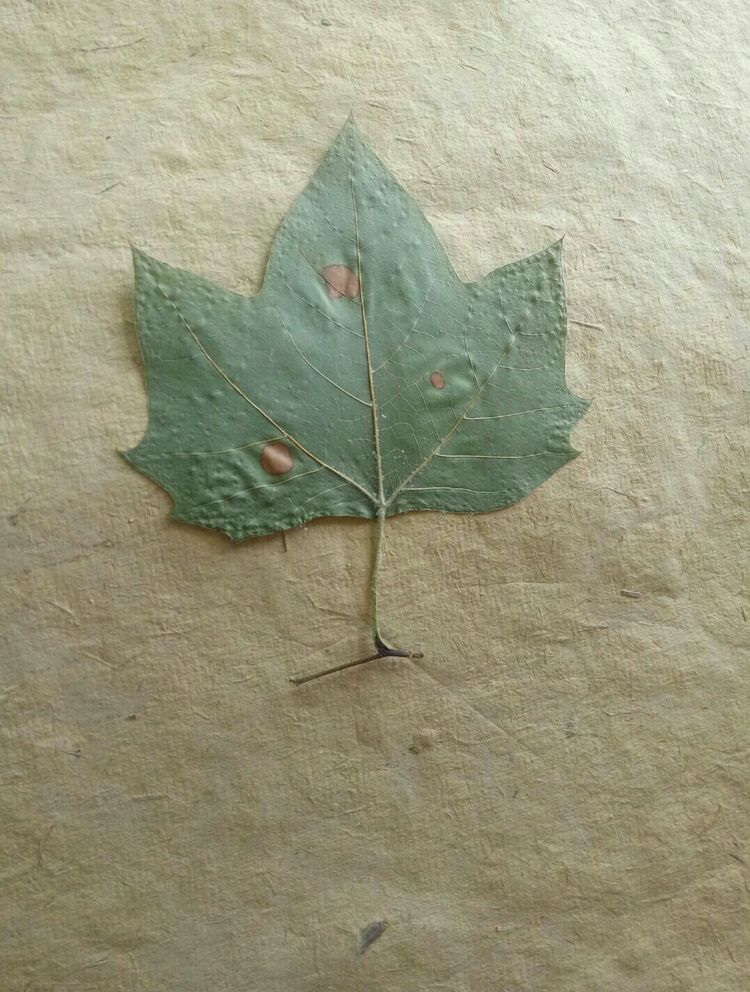
London plane tree anthrax
Symptoms:
Dark brown to black spots appear on both sides of the leaves, surrounded by yellowish-brown halos. Leaf spot disease of London plane trees is a common disease, generally occurring in June and causing leaf drop in September and October. In our county, recent continuous rainfall has caused some seedlings to turn yellow and lose their leaves.
Prevention and control methods:
Spray with a 1:1:100 Bordeaux mixture or a 500-fold dilution of 50% carbendazim wettable powder, once every 10 days, for 2-3 consecutive applications.
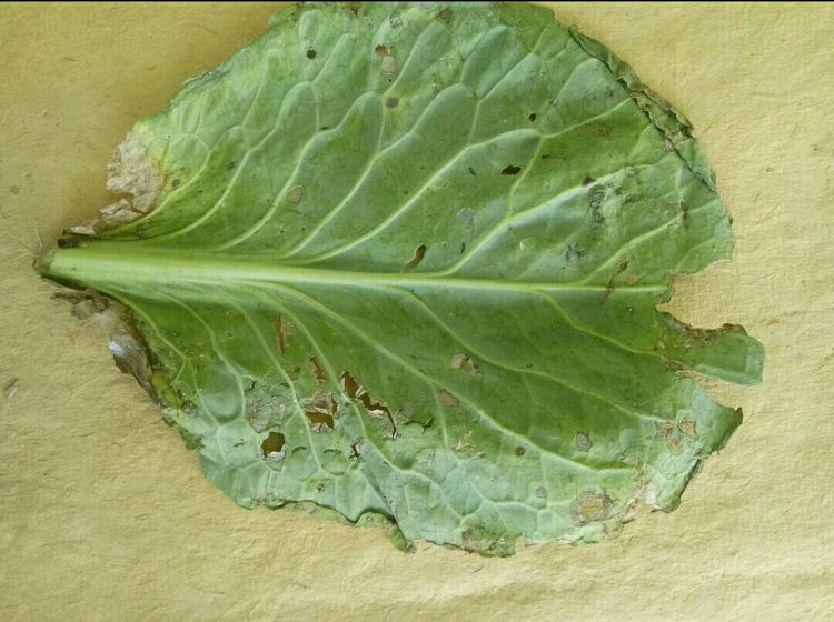
Black spot disease of cabbage
Pathogen, Latin name and classification: The pathogen is Alternaria brassicicola (Schweinitz) Wilts., a fungus belonging to the Deuteromycetes. [1]
Physiological and biochemical characteristics: The pathogen can develop in the range of 10-35℃, but the optimal temperature is relatively low, around 17℃.
Infection cycle: The pathogen overwinters in the soil, on diseased plant debris, and also on seed plants and seeds.
Conditions for onset
The disease does not occur when the relative humidity is below 80%. High humidity and heavy rainfall are key factors in the disease, as they weaken plant growth and exacerbate the disease.
Damaged parts: stems and leaves.
Harmful symptoms
It primarily affects the outer leaves and outer bulbous leaves, initially appearing as small, dark brown spots that expand into grayish-brown circular lesions 5-30 mm in diameter. The lesions show concentric rings, though these are not very distinct. Under high humidity, a dense black mold grows on the lesions. In severe cases, the lesions on the leaves often merge into large patches, causing the leaves to yellow and wither prematurely. On the stems and petioles, the lesions are longitudinal stripes and develop black mold.
Prevention and control methods
1. Use disease-free seeds. Generally, seeds should be disinfected by mixing them with 0.3% of 50% thiram or 50% iprodione.
2. Rotate crops between severely diseased areas and non-cruciferous vegetables for more than two years.
3. Apply sufficient manure and increase the application of phosphorus and potassium fertilizers. Apply topdressing and irrigation in a timely manner, and drain waterlogged fields after rain.
4. Remove diseased leaves promptly to reduce the source of infection. Clean the field after harvest and deeply till the soil.
5. In the early stage of the disease, timely chemical control can be carried out. 50% iprodione at 1500 times dilution, or 40% thiophanate-methyl at 400 times dilution, or 50% thiophanate-methyl at 800 times dilution, or 80% chlorpyrifos at 600 times dilution, or 75% chlorothalonil at 500 times dilution, or 40% captan at 400 times dilution.
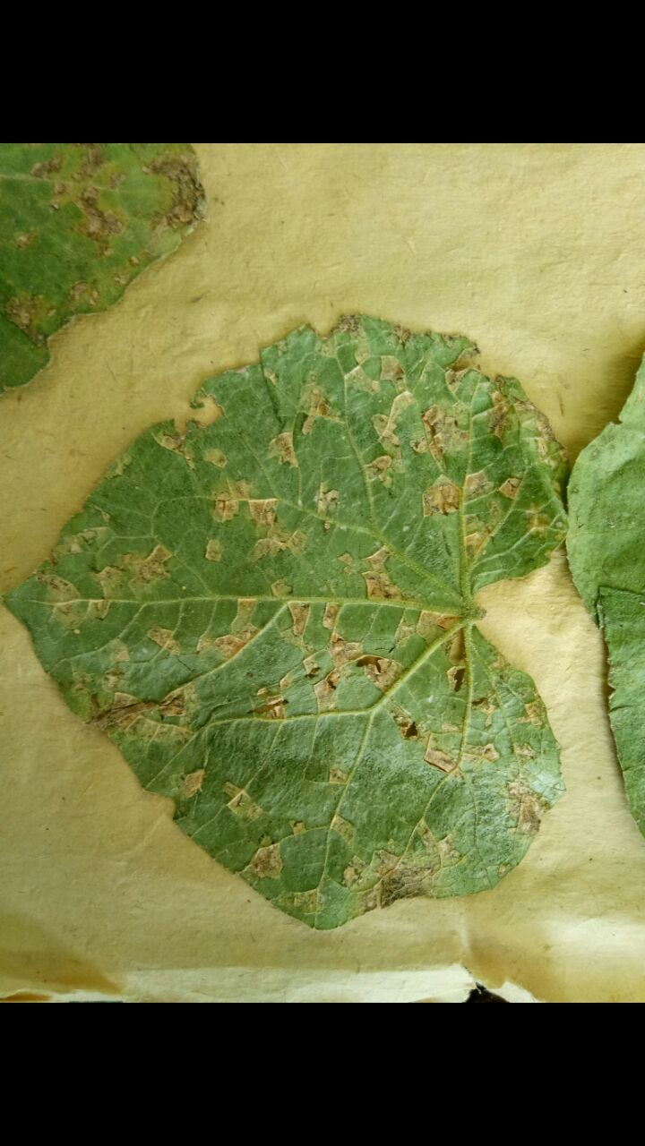
Cucumber angular leaf spot
Cucumber angular leaf spot is one of the important diseases affecting cucumbers. It often occurs in the field alongside cucumber downy mildew, with similar lesions that can sometimes be confused. However, in the early stages, cucumber downy mildew produces several polygonal, water-soaked lesions on the underside of the leaves, while bacterial angular leaf spot produces needle-like, water-soaked lesions on the underside of the leaves, often with dozens of lesions appearing simultaneously. The disease spreads less rapidly than downy mildew and has a less severe impact on cucumber growth.
Symptoms
Initially, tiny brownish spots appear on the true leaves. These spots gradually enlarge, turning yellowish-brown, and forming polygonal lesions about 3mm in diameter, surrounded by the leaf veins. At this stage, the area around the lesions turns yellow, forming a yellow halo. Then, the lesions gradually turn white, becoming brittle and easily broken.
pathogen
The pathogen Pseudomonas enters the plant through stomata and wounds on leaves and fruits, and is often spread by rain splashes.
Disease pattern
The pathogen overwinters inside or outside the seed, or in the soil with diseased plant debris, becoming the primary source of infection the following year. The seed-carrying rate is 2-3%. The pathogen enters through wounds on leaves or fruit vines, or through natural openings, penetrating the endosperm tissue or the outer cortex of the embryonic root, resulting in seed-borne infection. Additionally, diseased fruit can come into contact with contaminated seeds during harvesting, leading to external seed-borne infection. The pathogen can survive inside the seed for up to one year, and on diseased plant debris in the soil for 3-4 months. In production, if infected seeds are sown, the cotyledons will develop disease after emergence. The pathogen multiplies between cells. In greenhouses, the bacterial ooze from diseased parts of cucumbers spreads through the dripping of water droplets from the greenhouse roof, condensation, and leaf margins, causing repeated infections. In open-field cucumbers, disease first appears after the seedling stage ends, with the arrival of the rainy season and the start of field irrigation. The pathogen gradually spreads via airflow or rainwater, continuing until the peak fruiting period, after which the disease subsides as temperatures drop. The disease develops at temperatures between 10 and 30°C, with an optimal temperature of 24-28°C and a suitable relative humidity of over 70%. Low temperatures and high humidity in plastic greenhouses favor the disease. Lesion size is related to humidity: lesions are large and typical when nighttime saturated humidity exceeds 6 hours; lesions are small when humidity is below 85% or when saturated humidity lasts less than 3 hours; large diurnal temperature differences and heavy, prolonged dew formation exacerbate the disease. The day after watering in the field, numerous water-soaked lesions or bacterial ooze appear on the underside of leaves. Sometimes, even a small amount of inoculum can cause the disease to occur and spread.
Prevention and control methods
Bacterial angular leaf spot of cucumber primarily affects the leaves, but also the fruit and stems. After infection, the leaves initially show pale green, water-soaked spots, gradually turning yellowish-brown, light brown, pale yellow, grayish-white, and finally white. Due to the restriction of leaf veins, the lesions on the underside of the leaf are polygonal. When moist, the lesions on the underside of the leaf have a milky-white bacterial ooze; when dry, they appear as a thin white film or white powder, brittle and easily perforated.
When stems, petioles, and tendrils are infected, water-soaked spots appear, which extend longitudinally along the stem grooves into short strips. In high humidity, bacterial ooze may also appear. In severe cases, the diseased parts crack longitudinally, rot in a water-soaked manner, turn brown, and leave white marks on the surface after drying.
The lesions on the fruit initially appear as small, round, water-soaked spots. As they expand, they become irregular or contiguous lesions. The lesions crack and ooze white bacterial ooze. They are often accompanied by soft rot fungus infection, resulting in a yellowish-brown, water-soaked rot.
Prevention and control measures [2]:
Agricultural control: The disease is more likely to occur when fertilizer is insufficient and when plants are old. Seeds should be disinfected, and diseased leaves and plants should be removed and burned in time. [3]
2. Chemical control: Commonly used agents include 5% DT (copper oxychloride) at 500 times dilution, 72% agricultural streptomycin at 4000 times dilution, 77% copper oxychloride at 1200 times dilution, or 50 grams of 75% daconazole plus 15 grams of 72% agricultural streptomycin diluted in 15 kg of water for spraying. The above agents can be used alternately, spraying once every 4 to 5 days, for 3 to 4 consecutive sprays, and the control effect can reach more than 90%.
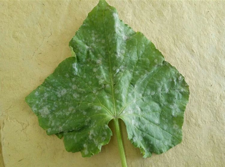
Cucumber powdery mildew
Symptoms of cucumber powdery mildew
Cucumber powdery mildew pathogen editing
*Erysiphe cichoracearum*, *Sphaerotheca fuliginea*, belongs to the Ascomycota and is a powdery mildew fungus belonging to the cucurbitaceae family. It is an obligate, weakly parasitic fungus, surviving only on living hosts. The conidiophores are cylindrical, and the conidia are oblong, colorless, and unicellular. The cleistothecia are brown, spherical, and contain one obovate ascus with eight elliptical ascospores. The optimal temperature for conidial production is 15–30°C, with a relative humidity above 75%.
Powdery mildew of cucumbers, commonly known as "white mold," most severely affects the leaves, followed by the petioles and stems, and generally does not harm the fruit. In the early stages of the disease, the upper surface of the leaves...
Powdery mildew on cucumbers causes small, nearly circular white powdery spots to appear on the leaves or undersides of the leaves. These spots gradually expand into large, indistinct white powdery areas, covering the entire leaf surface as if sprinkled with white powder. Wiping away the powder reveals that the leaves have lost their green color, turning yellow and brittle. In severe cases, the entire leaf surface is covered in powder, turning grayish-white, until the entire leaf dies. When powdery mildew infects petioles and young stems, the symptoms are similar to those on the leaves, but the lesions are smaller and contain less powder. Small yellow dots initially appear on the leaves, then expand into circular or oval lesions with a white powdery mold layer on the surface. Generally, there are more lesions on the lower leaves than the upper leaves, and more on the underside of the leaves than the upper side. The mold spots initially appear individually, but later merge into a large mold patch, sometimes covering the entire leaf, severely affecting photosynthesis, disrupting normal metabolism, causing premature aging, and resulting in yield loss.
Different varieties of flowers, trees, fruits and vegetables show significant differences in their resistance to powdery mildew. Selecting varieties with mild disease incidence or resistance is the most economical and effective way to control this disease.
(1) When purchasing seedlings, diseased plants must be strictly removed to eliminate the source of disease.
(2) When propagating, disease-free cuttings or root suckers should be taken as asexual propagation materials.
(3) When the melon seedlings are removed from the nursery, pesticides should be applied to prevent diseased seedlings from entering the new area.
1. Select disease-resistant varieties. Choose well-ventilated, loose, fertile soil with convenient irrigation and drainage for planting. Appropriately apply phosphorus and potassium fertilizers to prevent nutrient deficiency and premature aging, and enhance plant disease resistance. Do not water on cloudy days, and ventilate frequently on sunny days to reduce the relative humidity in greenhouses or polytunnels and prevent excessively high temperatures and stuffy conditions. In the early stages of cucumber powdery mildew or before disease onset, the main focus is on using protective agents to prevent infection and disease development.
2. When powdery mildew symptoms appear on the leaves in the field, use fast-acting treatments and mix them appropriately with a suitable amount of protective agent to prevent further damage and spread of the disease. 3. Strengthen management: Experience shows that when powdery mildew occurs, lightly watering between cucumber rows to increase air humidity, combined with spraying, can effectively control the disease. Other auxiliary measures include avoiding excessive application of nitrogen fertilizer, increasing the application of phosphorus and potassium fertilizers, and removing diseased plant debris after harvesting.